
FIGURE
Fig. 5
- ID
- ZDB-FIG-240524-23
- Publication
- Davison et al., 2024 - Tagging the tjp1a Gene in Zebrafish with Monomeric Red Fluorescent Protein Using Biotin Homology Arms
- Other Figures
- All Figure Page
- Back to All Figure Page
Fig. 5
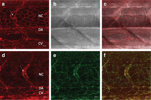
tjp1a-RFP embryos show localization of RFP to regions of cell?cell contact, and immunolabeling demonstrates that the RFP is colocalized with Tjp1a. (a?c) Confocal images of RFP in the tjp1a-RFP line in the trunk at 40 � with transmitted light in (b). Larva was 3 dpf. (d?f) Immunolabeling with anti-dsRed antibodies display a junctional pattern at 1 dpf at 63 � . (e) A similar pattern of labeling was observed with anti-Tjp1a antibodies. (f) Overlap of images in (d) and (e). NC, notochord; DA, dorsal aorta; CV, caudal vein, and the arrow indicates a segmental blood vessel. |
Expression Data
Expression Detail
Antibody Labeling
Phenotype Data
Phenotype Detail
Acknowledgments
This image is the copyrighted work of the attributed author or publisher, and
ZFIN has permission only to display this image to its users.
Additional permissions should be obtained from the applicable author or publisher of the image.
Full text @ Zebrafish