
Fig. 4
- ID
- ZDB-FIG-240524-22
- Publication
- Davison et al., 2024 - Tagging the tjp1a Gene in Zebrafish with Monomeric Red Fluorescent Protein Using Biotin Homology Arms
- Other Figures
- All Figure Page
- Back to All Figure Page
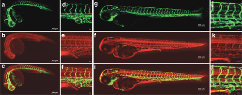
tjp1a-mRFP embryos show localization of mRFP to regions of cell?cell contact. Confocal images of mRFP (b, e, h, k), eGFP from fli1-egfpy1 (a, d, g, j), and merged (c, f, I, l) channels. (a?f) 1 dpf tjp1a-mRFPis86; fli1-egfpy1 embryos imaged with a 5x (a?c) and 20x (d?f) objective. (g?l) 3 dpf tjp1a-mRFP is86; fli1-egfpy1 embryos imaged with a 10x (g?i) and 20x (j?l) objective. All 20 � images are centered around the urogenital opening. eGFP from fli1-egfpn1 is expressed in blood vessels and mRFP from tjp1-mRFP is86 is likely present in tight junctions. mRFP was observed in blood vessels, the central nervous system, and the gut. |