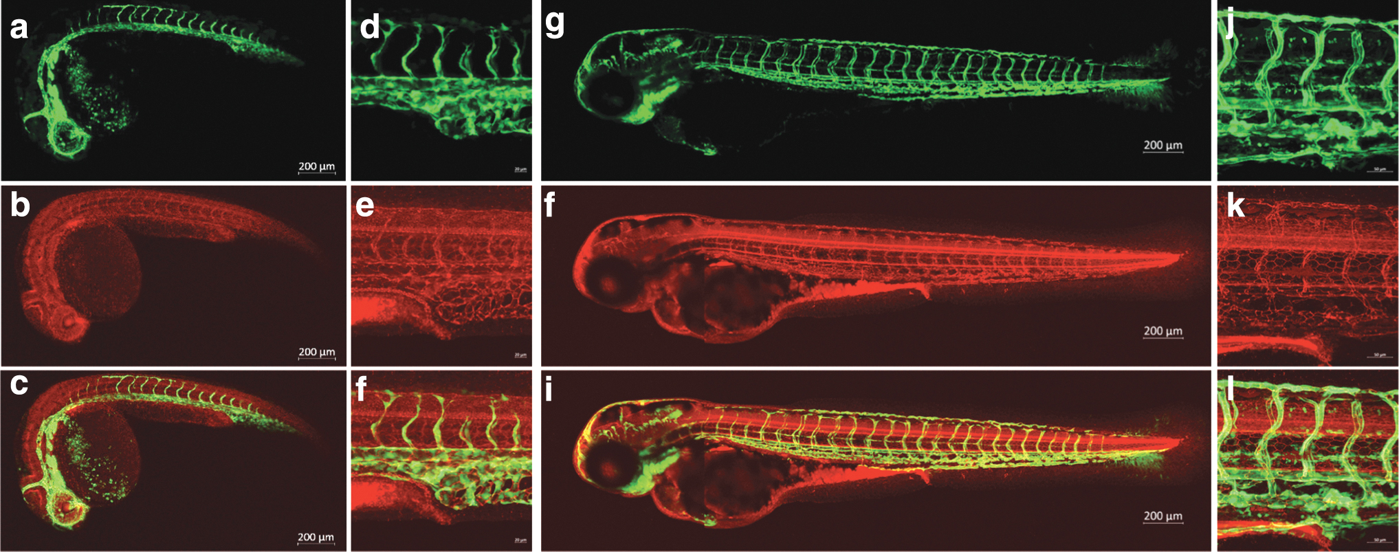
Fig. 4 tjp1a-mRFP embryos show localization of mRFP to regions of cell?cell contact. Confocal images of mRFP (b, e, h, k), eGFP from fli1-egfpy1 (a, d, g, j), and merged (c, f, I, l) channels. (a?f) 1 dpf tjp1a-mRFPis86; fli1-egfpy1 embryos imaged with a 5x (a?c) and 20x (d?f) objective. (g?l) 3 dpf tjp1a-mRFP is86; fli1-egfpy1 embryos imaged with a 10x (g?i) and 20x (j?l) objective. All 20 × images are centered around the urogenital opening. eGFP from fli1-egfpn1 is expressed in blood vessels and mRFP from tjp1-mRFP is86 is likely present in tight junctions. mRFP was observed in blood vessels, the central nervous system, and the gut.
Image
Figure Caption
Acknowledgments
This image is the copyrighted work of the attributed author or publisher, and
ZFIN has permission only to display this image to its users.
Additional permissions should be obtained from the applicable author or publisher of the image.
Full text @ Zebrafish

