
Fig. 6
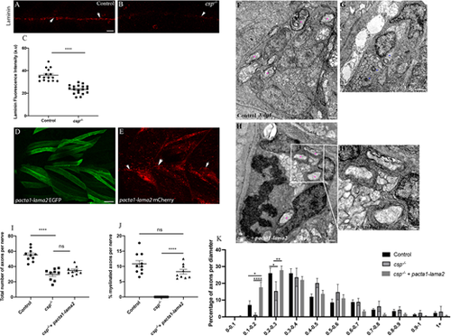
Laminin expression is significantly reduced in csp?/? and Laminin ?2 overexpression rescues radial sorting and myelination defects in csp?/? embryos. (A,B) Laminin expression in control (A) and csp?/? (B) embryos at 48 hpf showing the PLLn nerve (arrows). Scale bar: 20 ?m. (C) Quantification of laminin fluorescence intensity along the PLLn in controls (average of 35.95�1.39, n=15 embryos) and csp?/? (average of 23.37�0.90, n=18 embryos) embryos at 48 hpf (****P?0.0001), a.u., arbitrary unit. (D) Lateral view of EGFP expression in muscles surrounding the PLLn following pacta1-lama2 injection. Scale bar: 20 ?m. (E) Lateral view of mCherry-tagged secreted laminin in muscles and within the PLLn (white arrows). (F-H?) TEM of a cross-section of the PLLn at 3 dpf in control (F), csp?/? (G) and csp?/?+pacta1-lama2 (H) embryos. Magenta asterisks indicate some large caliber myelinated axons; some are shown at higher magnification in H?. Scale bars: 0.5 ?m. (I) Quantification of the total number of axons per nerve at 3 dpf in controls (average of 55.20�2.56, ten nerves, n=6 embryos), csp?/? (average of 29.56�2.55, nine nerves, n=7 embryos) and csp?/?+pacta1-lama2 (average of 34.78�1.84, nine nerves, n=7 embryos) (****P?0.0001; ns, P=0.1190). (J) Quantification of the percentage of myelinated axons relative to the total number of axons per nerve at 3 dpf in controls (average of 10.82�1.085), csp?/? (average of 0�0) and csp?/?+pacta1-lama2 (average of 8.28�0.77) (****P?0.0001; ns, P=0.075). (K) Graph representing the distribution of axons relative to their diameter with 0.1 �m bin width at 3 dpf in controls, csp?/? and csp?/? injected with pacta1-lama2 (*P=0.033 for 0.1-0.2; *P=0.04 for 0.2-0.3; **P=0.005; ****P<0.0001). |