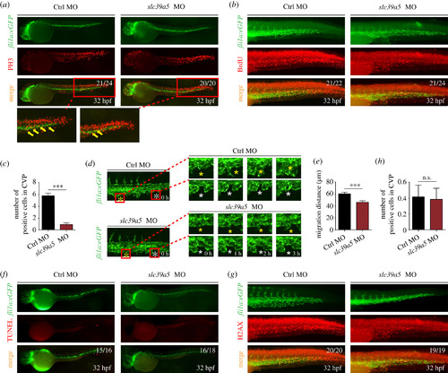
slc39a5 morphants have impaired endothelial cell proliferation and migration in the CVP region. (a?b) PH3 (a) and BrdU (b) immunostaining of control and slc39a5 morphant Tg(fli1a:eGFP) embryos, with magnified views of the CVP showing co-localization of GFP fluorescence and PH3 immunoreactivity (arrows). (c) Summary of BrdU-positive cells in the CVP of control and slc39a5 morphants. ***p < 0.001. (d) Representative sequential images of the CVP in control and slc39a5 morphant Tg(fli1a:eGFP) embryos. For each embryo, two separate endothelial cells are indicated with a white asterisk and a yellow asterisk. Note that 0 h corresponds to 32 hpf. (e) Summary of the migration distance of endothelial cells from 32 to 35 hpf in the CVP of control and slc39a5 morphant Tg(fli1a:eGFP) embryos. (f) Representative images of TUNEL staining in the CVP of control and slc39a5 morphant Tg(fli1a:eGFP) embryos. (g?h) Representative images (g) and summary (h) of H2AX immunostaining in the CVP of control and slc39a5 morphant Tg(fli1a:eGFP) embryos.
|
