
FIGURE
Fig. S6
- ID
- ZDB-FIG-160826-22
- Publication
- Ji et al., 2016 - Mutations in zebrafish pitx2 model congenital malformations in Axenfeld-Rieger syndrome but do not disrupt left-right placement of visceral organs
- Other Figures
- All Figure Page
- Back to All Figure Page
Fig. S6
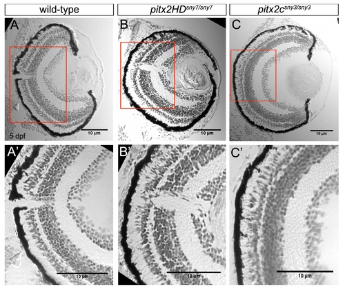
Retina layers are intact in pitx2 mutants at 5 dpf. (A-C) Cryosections of the eye from 5 dpf wild-type (A), pitx2HDsny7/sny7 (B) and pitx2csny3/sny3 (C) fish stained with crystal violet. (A′-C′) Enlarged view of boxes in A-C showing layers of the retina. Scale bars=10 �m. Supplementary material |
Expression Data
Expression Detail
Antibody Labeling
Phenotype Data
Phenotype Detail
Acknowledgments
This image is the copyrighted work of the attributed author or publisher, and
ZFIN has permission only to display this image to its users.
Additional permissions should be obtained from the applicable author or publisher of the image.
Reprinted from Developmental Biology, 416(1), Ji, Y., Buel, S.M., Amack, J.D., Mutations in zebrafish pitx2 model congenital malformations in Axenfeld-Rieger syndrome but do not disrupt left-right placement of visceral organs, 69-81, Copyright (2016) with permission from Elsevier. Full text @ Dev. Biol.