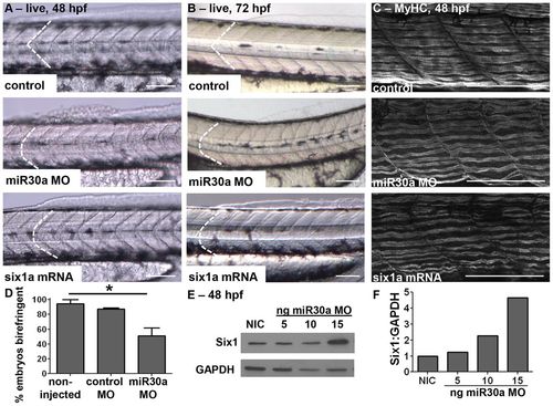
miR30a knockdown leads to abnormal somite and myosin fiber morphology, and to increased Six1 expression. Live images, anterior to the left, at (A) 48 hpf (control, n = 54/58; miR30a MO, n = 49/64; six1a mRNA, n = 36/45) and (B) 72 hpf (control, n = 75/75; miR30a MO, n = 30/40; six1a mRNA, n = 28/33) showing that miR30a knockdown (middle row) disrupts normal somite morphology to phenocopy six1a mRNA overexpression (bottom row). (C) Confocal images of MyHC stained with the A4.1025 antibody (control, n = 23/25; miR30a MO, n = 19/28; six1a mRNA, n = 11/20), and (D) loss of birefringence [mean±s.e.m.; non-injected control (NIC) n = 37 total embryos; control MO, n = 23; miR30a, n = 56; *P = 0.027, ANOVA with Bonferroni′s post-hoc test] demonstrate abnormal muscle fiber morphology upon miR30a loss at 48 hpf. (E,F) Increasing concentrations of miR30a-targeted MO result in a dose-dependent increase in (E) Six1 protein expression as visualized by western blot analysis at 48 hpf and (F) quantified following normalization to GAPDH. Scale bars: 100µm. For A, B, n = the number of embryos represented by each image/the total number of embryos analyzed.
|
