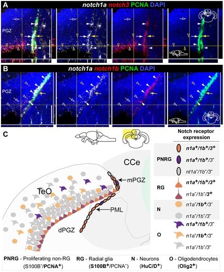
Overlapping and complementary notch1a/3 expression in the adult zebrafish optic tectum.
Confocal images showing localization of Notch receptor pairs by double FISH (white and red) and PCNA + proliferating cells (green). Cross-sections at the indicated level through the mesencephalon; tectal area shown in the micrographs is indicated in the cross section schematics. A, notch1a/3 and B, notch1a/1b expression domains in the dPGZ and mPGZ layers. Co-expression of these receptors both in PCNA + (filled white arrowheads) and PCNA cells (unfilled white arrowheads); notch3 -only cells in A and notch1b -only cells in B are indicated by unfilled yellow arrowheads; white arrows indicate notch1a + /notch3 - /PCNA + cells in A; a few PCNA cells are Notch receptor (filled yellow arrows); unfilled white arrows indicate cells positive for notch1a alone. C, Summary of Notch receptor expression pattern and cellular characteristics in the TeO. Abbreviations: Cce, corpus cerebelli; PGZ, periventricular gray zone of the optic tectum; dPGZ, deep layer of the PGZ; mPGZ, mitotic region of the PGZ; PML, posterior mesencephalic lamina; TeO, optic tectum. Scale bars = 50 μm.
|
