|
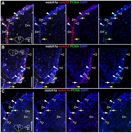
Overlapping and complementary Notch receptor expression in the dorsal telencephalon.
Confocal images showing localization of Notch receptors pairs by double FISH (white and red) and PCNA + proliferating cells (green); DAPI (blue) is used as nuclear counterstaining. Cross-sections at the indicated level through the telencephalon. Corresponding dorsal telencephalic areas represented in the micrographs are indicated in the cross section schematics of each panel. A, B, notch1a/3 co-expression in Dm and Dl, respectively. C, notch1a/b co-expression in Dm. These receptors are co-expressed both in PCNA + (filled white arrowheads) and PCNA - (unfilled white arrowheads) cells; within the PCNA + population, few cells are notch1a - /notch3 + (filled yellow arrowheads) while others are negative for both receptors (filled yellow arrows); within the PCNA - population, some cells express either notch3 or notch1b but not notch1a alone (unfilled yellow arrowheads). Scale bars = 50 μm.
|