
Fig. 3
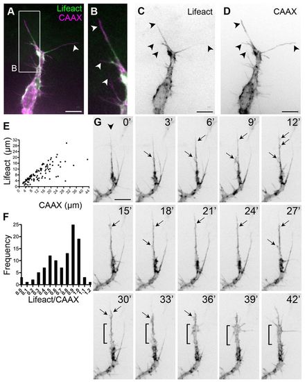
Dynamic F-actin localisation in filopodia. (A-D) Sprouting front of an ISV from a 30 hpf Tg(Fli1ep:Lifeact-EGFP); Tg(Kdr-l:ras-Cherry)s916 zebrafish embryo. Arrowheads indicate filopodia. (E) Length of the Lifeact signal in a CAAX-labelled filopodium. n=108 filopodia. (F) Frequency distribution of Lifeact/CAAX filopodia length. n=108 filopodia. (G) Dynamics of F-actin in a sprouting ISV from Tg(Fli1ep:Lifeact-EGFP) embryos. Lamellipodia-like structures (arrows) emanate from the most persistent filopodium (arrowhead) and give rise to a rapid expansion in vessel volume (from 30 to 42 minutes, bracket). Scale bars: 10 μm. |