FIGURE
Fig. S6
Fig. S6
|
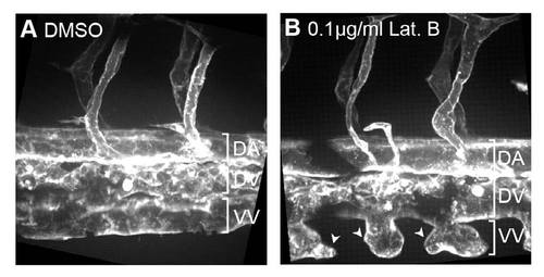
Endothelial cells of vein plexus are dependent on filopodia for anastomosis. Tg(Kdr-l:ras-Cherry)s916 embryos were treated with (A) 0.4% DMSO or (B) 0.1 μg/ml Lat. B from 22 hpf and imaged at 55 hpf. Vascular sprouts of the vein plexus become globular in morphology and fail to undergo anastomosis to form the ventral vein. DA, dorsal aorta; DV, dorsal vein; VV, ventral vein. Scale bars: 10 μm. |
Expression Data
Expression Detail
Antibody Labeling
Phenotype Data
Phenotype Detail
Acknowledgments
This image is the copyrighted work of the attributed author or publisher, and
ZFIN has permission only to display this image to its users.
Additional permissions should be obtained from the applicable author or publisher of the image.
Full text @ Development

