FIGURE
Fig. S2
Fig. S2
|
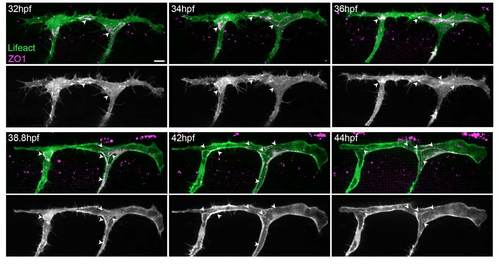
Actin filaments form at cell junctions. Still images from a movie of an embryo with clonal expression of Lifeact-EGFP and mCherry-hZO1 in ISVs and DLAV. F-actin (Lifeact) is formed at ZO1-positive cell junctions (arrowheads) and undergoes similar rearrangements as ZO1 as the ISV and DLAV become lumenised. Scale bar: 10 μm. |
Expression Data
Expression Detail
Antibody Labeling
Phenotype Data
Phenotype Detail
Acknowledgments
This image is the copyrighted work of the attributed author or publisher, and
ZFIN has permission only to display this image to its users.
Additional permissions should be obtained from the applicable author or publisher of the image.
Full text @ Development

