
Fig. S2
- ID
- ZDB-FIG-111031-9
- Publication
- Kroehne et al., 2011 - Regeneration of the adult zebrafish brain from neurogenic radial glia-type progenitors
- Other Figures
- All Figure Page
- Back to All Figure Page
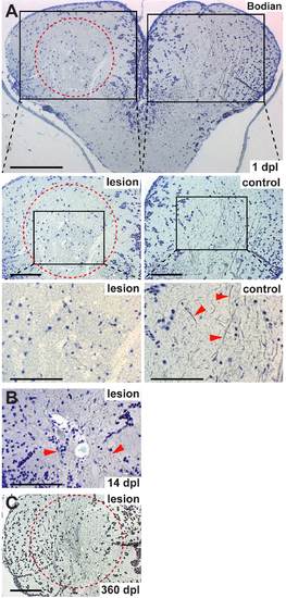
Neuronal processes get destroyed by the stab lesion, but are re-established 360 dpl. (A) Thick lateral axon bundles (arrowheads) are largely destroyed by the stab lesion, as seen in Bodian silver stainings of neuronal processes 1 dpl. (B) Already by 14 dpl neuronal processes re-appear within the lesion site (arrowheads), although the number and extent is still reduced compared with unlesioned telencephali. (C) By 360 dpl the number and thickness of the axon bundles is indistinguishable between lesioned (dashed circle) and unlesioned hemispheres (compare with A). Scale bars: 200 μm in A (low magnification); 100 μm in A (middle magnifications); 50 μm in A (high magnifications); 100 μm in B and C. All panels show 1 μm paraffin sections. Dashed outlines represent the lesion canal. |