
Fig. S7
- ID
- ZDB-FIG-111031-14
- Publication
- Kroehne et al., 2011 - Regeneration of the adult zebrafish brain from neurogenic radial glia-type progenitors
- Other Figures
- All Figure Page
- Back to All Figure Page
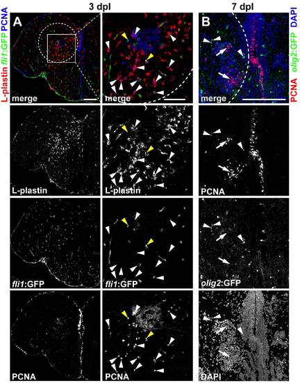
Most cells proliferating in the parenchyma are leukocytes. (A) At 3 dpl a sub-population of PCNA+ (blue), proliferating cells are found within and close to the lesion canal in the parenchyma. In the same area many ectopic L-plastin+ leukocytes (red) are located (see also Fig. 2B for comparison with the control hemisphere). Single confocal sections (inset) of 14 μm cryosections show that the great majority of PCNA+ nuclei in the parenchyma belong to L-plastin+ leukocytes (white arrowheads). Only a few PCNA+ nuclei are located within fli1-EGFP+ endothelial cells (green, yellow arrowheads). (B) 7 dpl olig2+:GFP OPCs (green) in the parenchyma are found to express PCNA (red, arrowheads), but many more proliferating cells in the parenchyma do not express olig2: GFP(arrows), as shown in single confocal sections. Scale bars: 100 μm in A, 50 μm in inset; 100 μm in B. Dashed outlines represent the lesion canal. |