
Fig. 7
- ID
- ZDB-FIG-111031-7
- Publication
- Kroehne et al., 2011 - Regeneration of the adult zebrafish brain from neurogenic radial glia-type progenitors
- Other Figures
- All Figure Page
- Back to All Figure Page
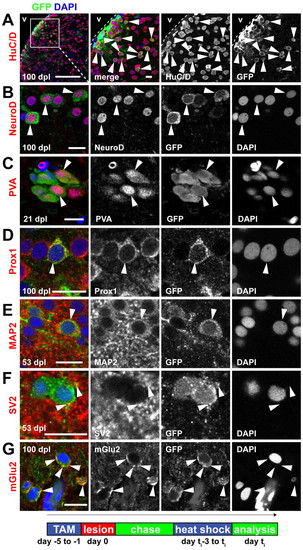
Radial glia-derived neurons are maintained long term, show a correct regional identity and display features of mature functional neurons. (A) Many recombined GFP+(green)/HuC/D+(red) neurons (arrowheads) are maintained in the parenchyma and in the PVZ until 100 dpl. (B-G) Examples of radial glia-derived (GFP+, green) neurons in the dorsal parenchyma that express: (B) the dorsal marker NeuroD1 (arrowheads, red) at 100 dpl; (C) the mature interneuron and regional marker parvalbumin (arrowheads, PVA, red) at 21 dpl; (D) the regional marker Prox1 (arrowhead, red) at 100 dpl; (E) the mature neuronal marker MAP2a+b (arrowhead, MAP2, red) at 53 dpl; (F) the synaptic vesicle marker SV2 (arrowheads, SV2, red) at 53 dpl; and (G) the synaptic marker metabotropic glutamate receptor 2 (arrowheads, mGlu2, red) at 100 dpl. Scale bars: 100 μm in A overview; 10 μm in A inset; in 10 μm in B-G. v, ventricle. Timeline shows experimental procedures. |