
Fig. 8
- ID
- ZDB-FIG-110920-76
- Publication
- Eames et al., 2011 - Mutations in fam20b and xylt1 Reveal That Cartilage Matrix Controls Timing of Endochondral Ossification by Inhibiting Chondrocyte Maturation
- Other Figures
- All Figure Page
- Back to All Figure Page
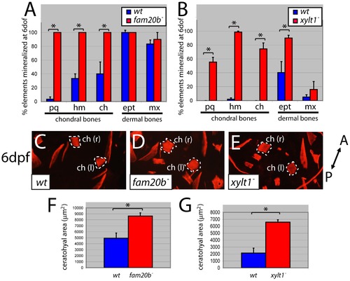
fam20b and xylt1 mutants have increased perichondral bone. At 6 dpf, Alizarin red staining (see Figure 1C?1G) was visible more often in skeletal elements of fam20bb1127 (A) and xylt1b1128 (B) mutants than in wild types. Statistically significant (*, p<0.05) increases, however, were observed in more chondral bones (pq, hm, ch) than dermal bones (ept, mx). Quantitative analyses of the sum of bone areas in left and right ceratohyals (ch(l) and ch(r), dashed outlines) in whole-mount, ventral images of live Alizarin red fluorescence (C?E) revealed statistically significant (*, p<0.05) increases in fam20bb1127 (F) and xylt1b1128 (G) mutants, compared to wild-type siblings at 6 dpf. Abbreviations: A = anterior; ch = ceratohyal; ept = entopterygoid; hm = hyomandibular; l = left; mx = maxilla; pq = palatoquadrate; P = posterior; r = right. |
| Fish: | |
|---|---|
| Observed In: | |
| Stage: | Day 6 |