Fig. 22
- ID
- ZDB-FIG-091217-16
- Publication
- Parichy et al., 2009 - Normal table of postembryonic zebrafish development: Staging by externally visible anatomy of the living fish
- Other Figures
-
- Fig. 1
- Fig. 2
- Fig. 5
- Fig. 6
- Fig. 8
- Fig. 10
- Fig. 11
- Fig. 13
- Fig. 14
- Fig. 16
- Fig. 17
- Fig. 18
- Fig. 19
- Fig. 21
- Fig. 22
- Fig. 23
- Fig. 24
- Fig. 25
- Fig. 26
- Fig. 27
- Fig. 28
- Fig. 32
- Fig. 33
- Fig. 34
- Fig. 35
- Fig. 36
- Fig. 37
- Fig. 38
- Fig. 39
- Fig. 40
- Fig. 41
- Fig. 42
- Fig. 43
- Fig. 44
- Fig. 45
- Fig. 46
- Fig. 47
- Fig. 48
- Fig. 49
- Fig. 50
- Fig. 51
- Fig. 52
- Fig. 53
- Fig. 54
- Fig. 55
- Fig. 56
- Fig. 57
- All Figure Page
- Back to All Figure Page
|
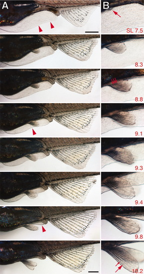
Pelvic fin development and fin fold minor lobe regression. Shown is a single individual (standard length [SL] at lower right). A: Low magnification views showing minor fin fold (arrowheads), vent, and anal fin. Subtle resorption of the minor fin fold is first apparent in this individual at ∼9.1 mm SL (arrowhead), and is clearly underway by 9.3 mm SL, when distal tips of the pelvic fins eclipse the edge of the fin fold; only small remnants of fin fold are present anterior to the vent at 9.8 mm SL (arrowhead) and 10.2 mm SL. B: Details of developing pelvic fin bud, first apparent in this individal at 7.5 mm SL (arrow). The bud extends from the body wall (8.3 mm SL), and three fin rays each comprising single developing segments are apparent by 8.8 mm SL (arrows). Second segments (large arrow) and the first joint between segments (small arrow) are clearly visible by 10.2 mm SL. Images shown are at decreasing magnifications. Scale bars = 7.5 and 10.2, 0.5 mm. |

