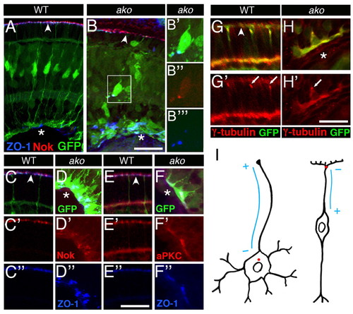
Apical polarity determinants are mislocalized in Müller glia. Transverse cryosections through wild-type or ako mutant retinae at 3 dpf. Sections are stained with antibodies against Nok, ZO-1, aPKC or γ-tubulin as indicated. The expression of the Tg(gfap:GFP) transgene marks Müller glia (green). (A,B) Nok (red) and ZO-1 (blue) localization in wild-type (A) and ako mutant (B) Müller glia. (B′-B″′) Detail of image shown in B; red, green and far red (in blue) channels are shown separately. (C-F″) Higher magnifications of Nok (red), Has/aPKC (red) and ZO-1 (blue) staining patterns. Retinae of mutant (D-D″,F-F″) and wild-type (C-C″,E-E″) siblings are shown next to each other. Nok (C′,D′) and aPKC (E′,F′) proteins colocalize with ZO-1 (bottom row of images), both in apical processes of wild-type Müller glia and in displaced glia of ako mutant eyes. Each column presents images obtained from a single section. Red and far red (in blue) channels are shown separately. (G-H′) Centrosome localization in Müller glia. Transverse cryosections through the retina stained with an antibody against γ-tubulin (red) to visualize basal bodies (arrows). (G′,H′) Red channel only. (I) Schematic drawing of a neuron (left) and a Müller cell (right) indicating the position of the centrosome (red dot) and the presumptive polarity of microtubules (blue lines). Asterisks indicate the vitreal (basal) surface of the retina; arrowheads indicate the OLM. Scale bars: 25 μm in A,B; 15 μm in C-F″; 10 μm in G-H′.
|

