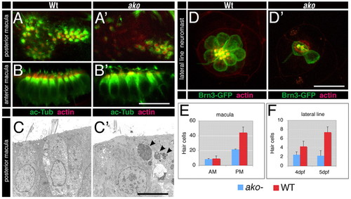
ale oko hair cell phenotype. (A-B′) Whole-mount staining of ear sensory maculae with anti-acetylated-α-tubulin antibody (green) and phalloidin (red, actin) to visualize hair cells and their stereocilia, respectively. Posterior (A′) and anterior (B′) maculae of ako mutants are shown. (C,C′) Electron micrographs of auditory hair cells in the posterior macula of wild-type (C) and ako mutant (C′) zebrafish larvae. Cellular debris (arrowheads) is present in the mutant tissue. (D,D′) In the ako mutant lateral line (D′), few hair cells survive by 5 dpf compared with the wild type (D). (E) Quantitation of hair cell numbers in the maculae of wild-type and akojj50 animals at 5 dpf. The average numbers of hair cells in the anterior (AM) and the posterior (PM) maculae are provided (n=6). For posterior macula, P<0.01 (Student′s t-test). (F) The average number of hair cells per lateral line neuromast in wild-type and akojj50 animals (n=6). At 4 and 5 dpf, P<0.0001 (Student′s t-test). A,A′,D,D′ show face views of the apical surface; in B-C′, apical is up. Scale bars: 5 μm in C,C′; 20 μm in A-B′,D,D′.
|
