|
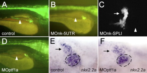
The development of the pancreatic ducts in nkx2.2a- and ptf1a-morphants. (A, B, D) Lateral views, anterior to the right. (C, E, F) Ventral views, anterior to the left. (A, E) Control embryos. (B, C) nkx2.2a-morphant larvae and (D, F) ptf1a-morphants. (A?D) GFP fluorescence in larvae of the nkx2.2(3.5):GFP line at 4 dpf. (A?D) The position of the intrapancreatic duct, which is lacking in panels B?D is indicated by an arrowhead. (C) Confocal image of a nkx2.2a-morphant larva where the extrapancreatic duct is marked by an arrow. A rudimentary duct is pointing in an anterior direction. (E, F) 48 hpf, putative precursors of the extrapancreatic duct express nkx2.2a (arrow) whereas there is a reduced number of nkx2.2a-expressing cells in the medial portion of the nkx2.2a expression domain (this area is marked by a dashed circle).
|