
|
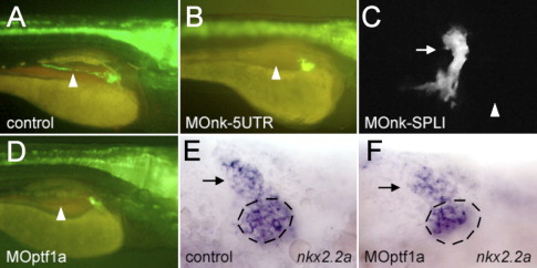
Fig. 7 The development of the pancreatic ducts in nkx2.2a- and ptf1a-morphants. (A, B, D) Lateral views, anterior to the right. (C, E, F) Ventral views, anterior to the left. (A, E) Control embryos. (B, C) nkx2.2a-morphant larvae and (D, F) ptf1a-morphants. (A?D) GFP fluorescence in larvae of the nkx2.2(3.5):GFP line at 4 dpf. (A?D) The position of the intrapancreatic duct, which is lacking in panels B?D is indicated by an arrowhead. (C) Confocal image of a nkx2.2a-morphant larva where the extrapancreatic duct is marked by an arrow. A rudimentary duct is pointing in an anterior direction. (E, F) 48 hpf, putative precursors of the extrapancreatic duct express nkx2.2a (arrow) whereas there is a reduced number of nkx2.2a-expressing cells in the medial portion of the nkx2.2a expression domain (this area is marked by a dashed circle).
Reprinted from Developmental Biology, 304(2), Pauls, S., Zecchin, E., Tiso, N., Bortolussi, M., and Argenton, F., Function and regulation of zebrafish nkx2.2a during development of pancreatic islet and ducts, 875-890, Copyright (2007) with permission from Elsevier. Full text @ Dev. Biol.