
Fig. 5
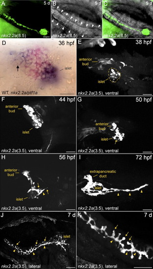
Confocal analysis of pancreatic duct morphogenesis in fixed nkx2.2a:GFP transgenic zebrafish. (A?C) Confocal images of a nkx2.2a(8.5) larva. (D) WT embryo. (E?K) Confocal images of nkx2.2a(3.5) embryos and larvae. (A?C) Lateral view, anterior to the right. (A) Fluorescent pancreatic duct (B) transmission, showing the acinar cells of the pancreatic tail (arrowheads) (C) overlay. (D) Ventral view, double in situ hybridization of nkx2.2a (blue) and ptf1a (red). nkx2.2a-positive cells of the anterior pancreatic bud are marked by an arrow. (E?I) Ventral views, anterior to the left. Different fixed individuals. The intrapancreatic duct is highlighted by arrowheads. The position of the pancreatic islet in panel E is indicated by a dashed circle. Arrows in panels G and H indicate filopodia. (J?K) Lateral view, anterior to the right. Arrows in panels J and K indicate first order branches. Scale bars: A?C and J 50 μm, D?I and K 25 μm. |
| Genes: | |
|---|---|
| Fish: | |
| Anatomical Term: | |
| Stage: | Prim-25 |
Reprinted from Developmental Biology, 304(2), Pauls, S., Zecchin, E., Tiso, N., Bortolussi, M., and Argenton, F., Function and regulation of zebrafish nkx2.2a during development of pancreatic islet and ducts, 875-890, Copyright (2007) with permission from Elsevier. Full text @ Dev. Biol.