
Fig. 10
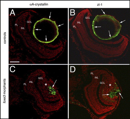
Lens cell marker expression in 7 dpf foxe3 morphants. Frozen sections from control (panels A and B) and morpholino-injected (panels C and D) embryos were immunolabeled for αA-crystallin and the zl-1 antigen at 7 dpf. In the controls, high levels of these proteins are detected in the peripheral lens cells (panels A and B, arrows). In the morphant lens, a subpopulation of cells express these proteins in an aberrant pattern (panels C and D, arrows). The asterisks in panels C and D mark the nucleated lens cell population. The scale bar (50 μm) in panel A applies to all panels. Abbreviations: PL, photoreceptor layer; INL, inner nuclear layer and GCL, ganglion cell layer. |
| Gene: | |
|---|---|
| Fish: | |
| Knockdown Reagent: | |
| Anatomical Term: | |
| Stage: | Days 7-13 |
| Fish: | |
|---|---|
| Knockdown Reagent: | |
| Observed In: | |
| Stage: | Days 7-13 |
Reprinted from Mechanisms of Development, 123(10), Shi, X., Luo, Y., Howley, S., Dzialo, A., Foley, S., Hyde, D.R., and Vihtelic, T.S., Zebrafish foxe3: Roles in ocular lens morphogenesis through interaction with pitx3, 761-782, Copyright (2006) with permission from Elsevier. Full text @ Mech. Dev.