
|
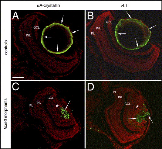
Fig. 10 Lens cell marker expression in 7 dpf foxe3 morphants. Frozen sections from control (panels A and B) and morpholino-injected (panels C and D) embryos were immunolabeled for αA-crystallin and the zl-1 antigen at 7 dpf. In the controls, high levels of these proteins are detected in the peripheral lens cells (panels A and B, arrows). In the morphant lens, a subpopulation of cells express these proteins in an aberrant pattern (panels C and D, arrows). The asterisks in panels C and D mark the nucleated lens cell population. The scale bar (50 μm) in panel A applies to all panels. Abbreviations: PL, photoreceptor layer; INL, inner nuclear layer and GCL, ganglion cell layer.
Reprinted from Mechanisms of Development, 123(10), Shi, X., Luo, Y., Howley, S., Dzialo, A., Foley, S., Hyde, D.R., and Vihtelic, T.S., Zebrafish foxe3: Roles in ocular lens morphogenesis through interaction with pitx3, 761-782, Copyright (2006) with permission from Elsevier. Full text @ Mech. Dev.