
- Title
-
Disturbed intracellular folate homeostasis impairs autophagic flux and increases hepatocytic lipid accumulation
- Authors
- Chi, W.Y., Lee, G.H., Tang, M.J., Chen, B.H., Lin, W.L., Fu, T.F.
- Source
- Full text @ BMC Biol.

Folate and folate-mediated one-carbon metabolism (OCM). a Folate comprises a pteridine ring and a p-aminobenzoic acid with polyglutamyl residues attached to the carboxyl group of the benzene ring. The one-carbon unit attached to the N5 and/or N10 positions of the pteridine ring is in the oxidation states of either formate, formaldehyde, or methanol. b Folate, through folate-mediated one-carbon metabolism (OCM), provides its one-carbon unit for purine and thymidylate biosynthesis, amino acid metabolism, S-adenosylmethionine formation, and redox homeostasis. Abbreviations: DHFR, dihydrofolate reductase; MTHFD1, methylenetetrahydrofolate dehydrogenase 1; SHMT1, cytosolic serine hydroxymethyltransferase; MTHFS, 5,10-methenyltetrahydrofolate synthetase; MTHFR, methylenetetrahydrofolate reductase; TS, thymidylate synthase; MTR, 5-methyltetrahydrofolate-homocysteine methyltransferase; MAT, methionine adenosyl transferase; MT, methyltransferase; SAHH, S-adenosylhomocysteine hydrolase; SHMT2, mitochondrial serine hydroxymethyltransferase-2; MTHFD1L, mitochondrial 10-formy-THF synthetase; MTHFD2/2L, mitochondrial bi-functional enzyme (5,10-methylene-THF dehydrogenase/5,10-methenyl-THF cyclohydrolase); MFT, mitochondrial folate transporter; RFC, reduced folate carrier; PCFT, proton-couple folate transporter; FR, folate receptor |
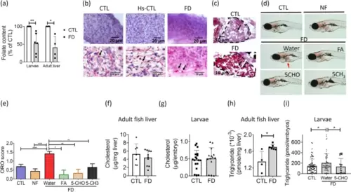
Increased lipid accumulation was found in FD zebrafish liver. The transgenic larvae and adult fish were induced for FD and examined for their hepatic lipid deposition. a The liver of larvae of 11 dpf and adult fish after completing FD-induction protocols were collected and subjected to microbiology assay. Data were collected from 6 and 4 independent experiments for larval liver and adult fish liver, respectively. b-c The cryosections prepared from adult fish liver (b) and whole larvae (c) were examined for lipid deposition (black arrows) with Oil Red O staining. d-e The hepatic lipid accumulation (red arrow) in larvae at 11 dpf, with/without FA, 5-CHO-THF, and 5-CH3-THF supplementation, were stained with Oil Red O (red arrow) (d) and scored (e) following the criteria based on the percentage of intensely stained area occupied in larval liver: less than 30% (1), between 30% to 70% (2), or more than 70% (3). Those showed no significant staining signal was scored (0). Data were collected from at least 3 independent experiments. f-i Hepatic cholesterol (f-g) and triglyceride (h-i) in adult fish liver (f and h) and larvae at 11 dpf (g and i) were measured with colorimetric/fluorometric Assay. Significant increase was found for the triglyceride levels in both adult fish liver and larval liver. Supplementing with 5-CHO-THF effectively prevented the increase of triglyceride in FD larvae. All the data presented are the averages of at least three independent trials with 10-40 larvae or 4-10 adult fish livers for each group. CTL, control (Tg-GGH/LR larvae without FD); FD, folate deficiency; FA, folic acid; 5-CHO, 5-formyl-THF; 5-CH3, 5-methyl-THF. Statistical data are shown in mean � SEM. * p<0.05, **, p <0.01; ****, p<0.0001 |
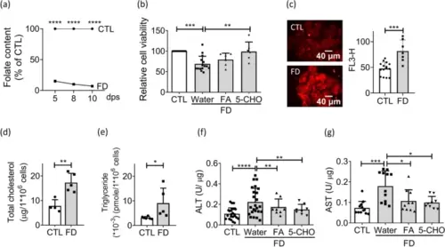
Increased lipid deposition was found in FD Huh7 cells. Huh7 cells were cultivated in FD medium and examined for cell viability and lipid content. a Huh7 cells cultivated in CTL (0.004 g/L folic acid) or folate-deficient (FD) medium were analyzed with microbiology assay for folate content. A more than 90% depletion of intracellular folate content was established at 10 day-post-seeding (dps) for the cells growing in FD medium. Presented are data collected from 3 and 4 independent experiments at 5 dps, and 8 and 10 dps, respectively. b Huh7 cells were cultivated in CTL or FD medium for 7 days (dps) before adding FA or 5-CHO-THF for rescue. Cell viability was examined on 8 dps. The decreased cell viability due to FD was successfully rescued by the presence of FA or 5-CHO-THF. Data were collected from at least 6 independent experiments. c Cells were stained with Nile red and analyzed for lipid content with direct visual inspection with fluorescence microscopy (left) and quantified by flow cytometry (right). Scale bars = 40 �m. Data were collected from at least 7 independent experiments. d-e The cholesterol (d) and triglyceride (e) levels of Huh7 cells at 8 dps was measured with colorimetric/fluorometric Assay. Data were collected from at least 6 independent experiments. f-g Cells cultivated in FD medium with/without FA or 5-CHO-THF supplementation were examined for ALT (f) and AST (g) content. The increase of both ALT and AST was effectively alleviated by supplementing with FA or 5-CHO-THF. Presented are the averaged results of at least 8 independent trials. CTL, control (cells without FD); FD, folate deficiency. Statistical data are shown in mean � SEM. * p<0.05, **, p <0.01; ***, p<0.001, ****, p<0.0001 |
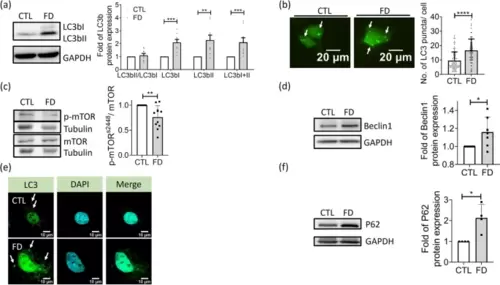
Impaired autophagic flux was found in FD Huh7 cells. Huh7 cells cultivated in CTL (0.004 g/L folic acid) or FD medium for 8 days were examined for the occurrence and flow of autophagy. a The expression of LC3b, the autophagic marker, was examined with Western blotting (left) and quantified (right). The increase of LC3bI and LC3bII signified an activated autophagy. Presented are the averaged results of 9 independent trials. b Cells transfected with plasmids encoding LC3-eGFP were cultivated in CTL (0.004 g/L folic acid) or folate deficient (FD) medium and examined for the green fluorescent puncta, which represent the LC3-containing autophagic vesicles. Scale bars = 20 �m. Data were collected from at least 3 independent experiments with the total sample number of 66-97 for each group. c-d Cells were examined for the expression of mTOR, p-mTORs2448 and Beclin1. Significantly decreased p-mTOR s2448/mTOR ratio and increased Beclin1 were observed in FD Huh7 cells, suggesting an activation of autophagy and formation of phagophore. Presented are data collected from 11 and 7 independent experiments for mTOR and Beclin1, respectively. e Huh7 cells were immuno-stained for LC3 distribution and examined with FV3000 confocal laser scanning microscopy. The increased relocation of dispersed LC3 puncta to cytoplasm (white arrows) was found in FD Huh7 cells. Scale bars = 10 �m. f Apparently increased P62 was observed in FD Huh7 cells, indicating an increased autophagosomes. Presented are the averaged results of four independent trials. CTL, control (cells without FD); FD, folate deficiency. Statistical data are shown in mean � SEM. * p<0.05, **, p <0.01; ***, p<0.001, ****, p<0.0001 |
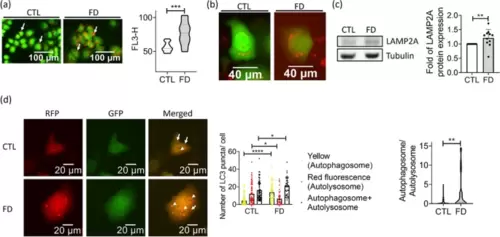
FD caused autophagosomes accumulation in Huh7 cells. Huh7 cells grown in FD medium for 5 days were stained with acridine orange (a) and LysoTracker (b) for acidic vesicles. Data were collected from at least 3 independent experiments. Cells were also subjected to Western blotting for LAMP2A, a lysosomal membrane protein promoting autophagic flux (c). Data were collected from 11 independent experiments. d Cells transfected with mRFP-GFP tandem fluorescence-tagged LC3 plasmid were examined for the presence of autophagosome (yellow fluorescent dots) and autolysosome (red fluorescent dots) (left). Increased autophagosomes and decreased autolysosomes were found in FD Huh7 cells (middle), leading to a significantly elevated autophagosome/autolysosome ratio (right). Scale bars = 20 �m. Presented are the averaged results of at least three independent trials. CTL, control (cells without FD); FD, folate deficiency. Statistical data are shown in mean � SEM. * p<0.05, **, p <0.01; ***, p<0.001, ****, p<0.0001 |
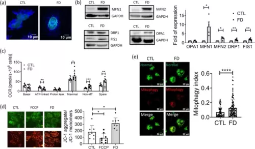
Disturbed mitochondrial activity and distribution were found in FD Huh7 cells. Huh7 cells cultivated in FD medium for 5 days were stained with MitoTracker for mitochondrial morphology (a) and subjected to Western blotting for characterizing the expression of mitochondrial fusion-fission proteins (b). Significant increase was found for both fusion (MFN1 and MFN2) and fission (DRP1 and FIS1) proteins, suggesting an increased mitochondrial dynamic in FD cells. Data were collected from at least 4 independent experiments. c Huh7 cells cultivated in CTL or FD medium for 8 to 9 days were subjected to Oroboros O2k for monitoring mitochondrial activity. Lowered ATP production and elevated non-mitochondria OCR and spare capacity were observed in Huh7 cells. Presented are data collected from at least 9 independent experiments. d Cells cultivated in CTL or FD medium for 8 days were stained with JC-1 (5,5',6,6'-tetrachloro-1,1',3,3'-tetraethylbenzimidazolylcarbocyanine iodide) for assessing mitochondrial membrane potential (MMP) changes. The increased ratio between aggregated and monomer mitochondria indicates a higher mitochondrial membrane potential in FD cells. Presented are data collected from at least 8 independent experiments (e) Huh7 cells were transfected with mt-Keima, a ratio-metric pH-sensitive fluorescent probe targeting the mitochondrial matrix, at 7 dps. In a neutral environment, mt-Keima predominantly emits a short wavelength (normal, 440 nm), displaying a green mitochondrial signal. Conversely, in an acidic environment, it primarily emits a long wavelength (mitophagy, 586 nm), generating a red lysosomal signal. The 'Mitophagy Index' was determined by calculating the ratio of 586 nm/ 440 nm fluorescence signals in Huh7 cells. Data were collected from at least 3 independent experiments with the total sample number of 90-97 for each group. Statistical data are shown in mean � SEM. CTL, control (cells without FD); FD, folate deficiency. FCCP, carbonylcyanide-p-trifluoromethoxyphenylhydrazone (a potent uncoupler of oxidative phosphorylation). Statistical data are shown in mean � SEM. * p<0.05, **, p <0.01, ****, p<0.0001 |
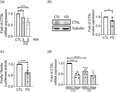
The expression of CTSL was down-regulated in FD Huh7 cells. a Huh7 cells cultivated in FD medium were harvested at 3-dps and 5-dps and characterized for cathepsin L expression with real-time PCR. Data were collected from at least 3 independent experiments. b The protein levels of cathepsin L, which were examined after cells being cultivated for 5 days, were significantly decreased in those grown in FD medium. The average of nine independent experiments. c Huh7 cells transfected with plasmids encompassing zebrafish cathepsin L promoter region (-2500 to +270) were harvested 3 days after transfection and subjected to promoter activity assay. The average of ten independent experiments. d Cells cultivated in medium containing rapamycin, a mTOR inhibitor, were examined for cathepsin L expression at 5-dps. The presence of rapamycin lowered cathepsin L mRNA levels in control groups, but did not cause further decrease in FD cells. Data were collected from at least 5 independent experiments. CTL, control (cells without FD); FD, folate deficiency; CTSL, cathepsin L. Statistical data are shown in mean � SEM. * p<0.05, **, p <0.01; ***, p<0.001, ****, p<0.0001 |

Inhibiting cathepsin L activity increased the number of acidic vesicles and lipid accumulation in Huh7 cells. a-b Huh7 cells grown in medium containing 40 �M cathepsin L inhibitor (CTSL-I) for 2 days were stained with acridine orange for acidic vesicles (a) and Nile red for lipid accumulation (b). Cells were imaged with fluorescence microscopy (left) and quantified for fluorescence intensity with flow cytometry (right). Increased numbers of red fluorescent puncta/intensity were found in FD cells, which were effectively prevented by folate supplementation. Data were collected from at least 6 independent experiments. c Cells transfected with hCTSL/eGFP/pCS2+ plasmids encoding recombinant human cathepsin L were subjected to Nile red staining for lipid deposition (left) and quantified with flow cytometry (middle). The expression of recombinant human cathepsin L was confirmed with Western blotting (right). Presented are the averaged results of at least five independent trials. CTL, control (cells without FD); FD, folate deficiency; CTSL, cathepsin L. Statistical data are shown in mean � SEM. * p<0.05, **, p <0.01; ***, p<0.001, ****, p<0.0001. Scale bars = 40 �m |

The folate deficiency caused by methotrexate altered autophagic activity and decreased cathepsin L expression in Huh7 cells. a Huh7 cells were examined for viability after cultivating in regular DMEM medium containing methotrexate (MTX) of indicated concentrations. An approximately 30% decrease in cell viability was observed in the presence of MTX at 1 �M and beyond. Data were collected from 5 independent experiments. b An almost complete depletion of intracellular folate content was reached after cells were cultivated in 5 �M MTX for 2 days. Data were collected from 6 independent experiments. c-e Cells grown in 5 �M MTX for 2 days were stained with Nile red for lipid deposition (c), transfected with plasmids encoding LC3-eGFP for characterizing autophagic activity (d) and subjected to Acridine orange staining for acidic vesicles (e). Increased lipid accumulation, autophagy and lysosomes/autolysosomes were apparent in MTX-treated cells. Data were collected from at least 3 independent experiments. f Cells were harvested 2 days after exposing to 5 �M MTX and examined for cathepsin L mRNA. A significant decrease in cathepsin L expression was found for MTX-treated cells. Presented are the averaged results of at least three independent trials. MTX, methotrexate. Statistical data are shown in mean � SEM. * p<0.05, **, p <0.01; ***, p<0.001 |
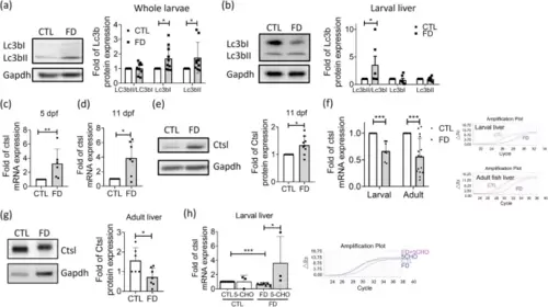
Up-/dysregulation of autophagy and down-regulation of cathepsin L expression also occurred in the liver of FD fish in vivo. a-b Cell lysates prepared from the whole larvae (a) and isolated liver from larvae of 11 dpf (b) were Western blotted for Lc3b and quantified with densitometry. Elevated Lc3bII/Lc3bI ratio was most apparent in FD larval liver, signifying an enhanced autophagosome formation, interrupted autophagosome-lysosome fusion, or lysosomal deregulation. Data were collected from at least 7 independent experiments. (c-d) Real-time PCR performed with the sample prepared from whole larvae of 5 dpf (c) and 11 dpf (d) revealed increased cathepsin L expression. Data were collected from at least 6 independent experiments. e Western blotting was conducted with the extract prepared from the whole larvae of 11 dpf for cathepsin L protein level, which revealed an increased expression for FD group. Data were collected from 9 independent experiments. f-g The liver-specific decrease in the expression of cathepsin L was observed both in the larvae of 11 dpf and adult fish of FD groups for both mRNA (f) and protein (g) levels. Data were collected from at least 6 independent experiments. h Larvae were induced for FD and supplementing with 1 mM 5-CHO-THF from 7 to 11 dpf before collected and examined for cathepsin L expression. Folate supplementation significantly increased the cathepsin L mRNA in FD larval liver. Presented are the averaged results of at least three independent trials with each of the sample prepared from 10-20 larvae. CTL, control (Tg-GGH/LR larvae without FD); FD, folate deficiency; 5-CHO, 5-formyl-THF. Statistical data are shown in mean � SEM. * p<0.05, **, p <0.01; ***, p<0.001 |
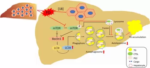
The prospective pathomechanism contributing to the FD-induced hepatic lipid accumulation. FD prevents mTOR phosphorylation, which enhances autophagic initiation. The decreased mTOR phosphorylation also down-regulates cathepsin L expression, which results in ineffective lysosomal activity and impaired autolysosome maturation, leading to disrupted autophagic flux and hepatic lipid accumulation |