
|
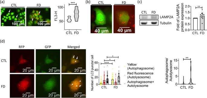
Fig. 5 FD caused autophagosomes accumulation in Huh7 cells. Huh7 cells grown in FD medium for 5 days were stained with acridine orange (a) and LysoTracker (b) for acidic vesicles. Data were collected from at least 3 independent experiments. Cells were also subjected to Western blotting for LAMP2A, a lysosomal membrane protein promoting autophagic flux (c). Data were collected from 11 independent experiments. d Cells transfected with mRFP-GFP tandem fluorescence-tagged LC3 plasmid were examined for the presence of autophagosome (yellow fluorescent dots) and autolysosome (red fluorescent dots) (left). Increased autophagosomes and decreased autolysosomes were found in FD Huh7 cells (middle), leading to a significantly elevated autophagosome/autolysosome ratio (right). Scale bars = 20 �m. Presented are the averaged results of at least three independent trials. CTL, control (cells without FD); FD, folate deficiency. Statistical data are shown in mean � SEM. * p<0.05, **, p <0.01; ***, p<0.001, ****, p<0.0001