
|
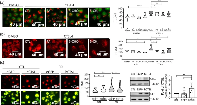
Fig. 8 Inhibiting cathepsin L activity increased the number of acidic vesicles and lipid accumulation in Huh7 cells. a-b Huh7 cells grown in medium containing 40 �M cathepsin L inhibitor (CTSL-I) for 2 days were stained with acridine orange for acidic vesicles (a) and Nile red for lipid accumulation (b). Cells were imaged with fluorescence microscopy (left) and quantified for fluorescence intensity with flow cytometry (right). Increased numbers of red fluorescent puncta/intensity were found in FD cells, which were effectively prevented by folate supplementation. Data were collected from at least 6 independent experiments. c Cells transfected with hCTSL/eGFP/pCS2+ plasmids encoding recombinant human cathepsin L were subjected to Nile red staining for lipid deposition (left) and quantified with flow cytometry (middle). The expression of recombinant human cathepsin L was confirmed with Western blotting (right). Presented are the averaged results of at least five independent trials. CTL, control (cells without FD); FD, folate deficiency; CTSL, cathepsin L. Statistical data are shown in mean � SEM. * p<0.05, **, p <0.01; ***, p<0.001, ****, p<0.0001. Scale bars = 40 �m