
Fig. 4
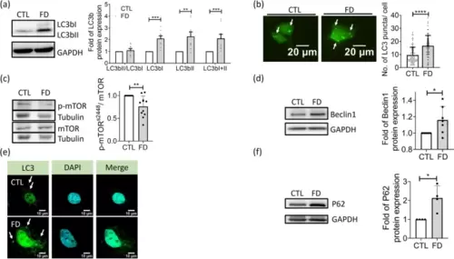
Impaired autophagic flux was found in FD Huh7 cells. Huh7 cells cultivated in CTL (0.004 g/L folic acid) or FD medium for 8 days were examined for the occurrence and flow of autophagy. a The expression of LC3b, the autophagic marker, was examined with Western blotting (left) and quantified (right). The increase of LC3bI and LC3bII signified an activated autophagy. Presented are the averaged results of 9 independent trials. b Cells transfected with plasmids encoding LC3-eGFP were cultivated in CTL (0.004 g/L folic acid) or folate deficient (FD) medium and examined for the green fluorescent puncta, which represent the LC3-containing autophagic vesicles. Scale bars = 20 �m. Data were collected from at least 3 independent experiments with the total sample number of 66-97 for each group. c-d Cells were examined for the expression of mTOR, p-mTORs2448 and Beclin1. Significantly decreased p-mTOR s2448/mTOR ratio and increased Beclin1 were observed in FD Huh7 cells, suggesting an activation of autophagy and formation of phagophore. Presented are data collected from 11 and 7 independent experiments for mTOR and Beclin1, respectively. e Huh7 cells were immuno-stained for LC3 distribution and examined with FV3000 confocal laser scanning microscopy. The increased relocation of dispersed LC3 puncta to cytoplasm (white arrows) was found in FD Huh7 cells. Scale bars = 10 �m. f Apparently increased P62 was observed in FD Huh7 cells, indicating an increased autophagosomes. Presented are the averaged results of four independent trials. CTL, control (cells without FD); FD, folate deficiency. Statistical data are shown in mean � SEM. * p<0.05, **, p <0.01; ***, p<0.001, ****, p<0.0001 |