- Title
-
aldh7a1 Regulates Eye and Limb Development in Zebrafish
- Authors
- Babcock, H.E., Dutta, S., Alur, R.P., Brocker, C., Vasiliou, V., Vitale, S., Abu-Asab, M., Brooks, B.P.
- Source
- Full text @ PLoS One
|
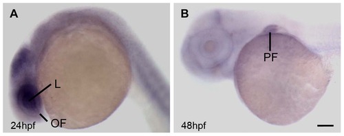
Expression pattern of aldh7a1 in zebrafish. Whole-mount in situ hybridization of aldh7a1 at (A) 24 hpf and (B) 48 hpf. L, lens; OF, optic fissure; PF, pectoral fin. Scale bar: 65 μm in A; 60 μm in B. |
|
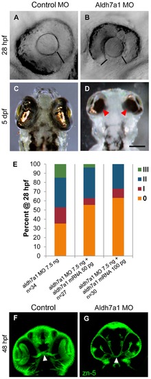
Aldh7a1 is important for optic fissure closure. (A?D) Injection of Aldh7a1 morpholino results in failure of optic fissure to close at 28 hpf and sustained at 5 dpf. (A) Eye of control morpholino (MO) injected embryo at 28 hpf, (B) Eye of aldh7a1 morphant at 28 hpf; (C) Ventral view of eye in control MO embryo at 5 dpf, (D) Ventral view of eye in embryos injected with 7.5 ng Aldh7a1 MO at 5 dpf, black bars indicate edges of optic fissures; (E) Bar graph demonstrate distribution of eye phenotypes 0, I, II, and III at 28 hpf following 7.5 ng Aldh7a1 MO injection followed by partial rescue of phenotype upon co-injection of two doses of aldh7a1 mRNA. All control MO injected embryos displayed ?0? phenotype. (F) zn-5 staining of control MO injected embryos at 48 hpf compared to (G) aldh7a1 morphant which displays optic nerve hypoplasia, arrow-heads indicate optic nerve. Scale bar: 65 μm in A,B; 125 μm in C,D; 75 μm in F,G. |
|
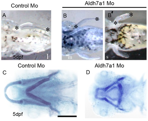
Aldh7a1 morpholino knockdown embryos show defects in pectoral fin and cartilage development. (A?B2) Fin phenotypes long (l), medium (m), and short (s) classified by length at 5 dpf, marked by black asterisks. All control MO embryos displayed ?Long? fins (A) and Aldh7a1MO injected embryos develop medium (B, 6%) or short (B2, 10%) fin. (C?D) Ventral view of Alcian blue staining of jaw- cartilages in control MO (C) and aldh7a1morphant (D) embryos. Scale bar: 350 μm in A?B2 125 μm in C,D. PHENOTYPE:
|
|
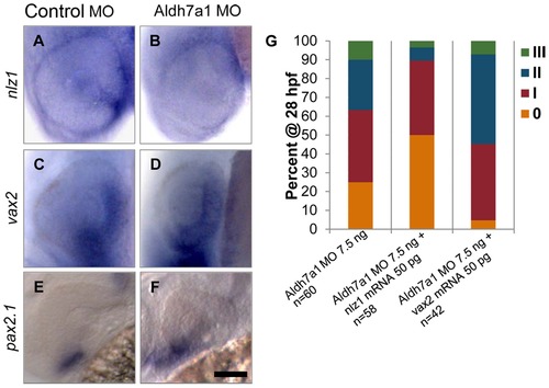
Expression pattern of genetic eye development markers in control and aldh7a1 morphant embryos. (A) Expression of nlz1 in optic fissure is down-regulated in (B) aldh7a1 morphant fish. vax2 and pax2.1 do not seem to show significant change in expression between control MO (C,E) and nlz1 morphant (D, F) fish. (G) Co-injection of nlz1 mRNA resulted in partial rescue of aldh7a1 MO phenotype, examined at 28 hpf, while co-injection of vax2 mRNA showed no change. Scale bar: 65 μm. EXPRESSION / LABELING:
|
|
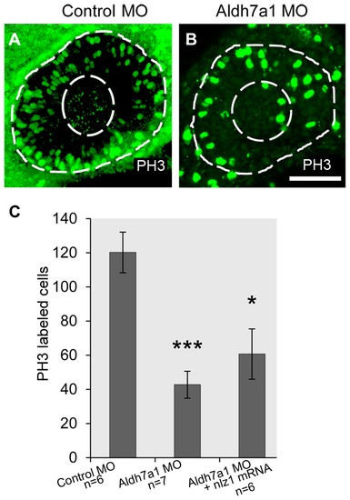
Aldh7a1 is required for retinal cell proliferation. (A?B) Dividing cells in developing eye were labeled with phosophohistone-3 antibody (H3P) in (A) Control MO and (B) alh7a1morphant embryos at 28 hpf. (A) and (B) are projection images of z-stacks through the depth of the eye. (C) Average number of dividing cells per eye quantified for control MO (n = 6), Nlz1 MO (n = 7), nlz1 mRNA (n = 6) rescued Statistical significance indicated above columns *P<0.05, **P<0.01, ***P<0.0001. Scale bar: 65 μm. PHENOTYPE:
|
|
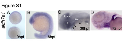
aldh7a1 expression. Whole-mount in situ hybridization of shows expression of aldh7a1 at (A) 9hpf, (B) 18hpf,(C) 36hpf, and (D) 72hpf.e,eye; t, tectum; ce,cerebellum; ov, otic vesicle; fb, fin bud; pf, pectoral fin. Arrow head indicates pharyngeal arches. EXPRESSION / LABELING:
|
|
Grades of severity for eye development. Classification of phenotype severity following morpholino injections. Four grades of coloboma (0, least severe; I, II,and III, most severe) classified by distance between edges of optic fissure at ~28 hpf, marked by white asterisks. Scale bar: 65 μm |
|
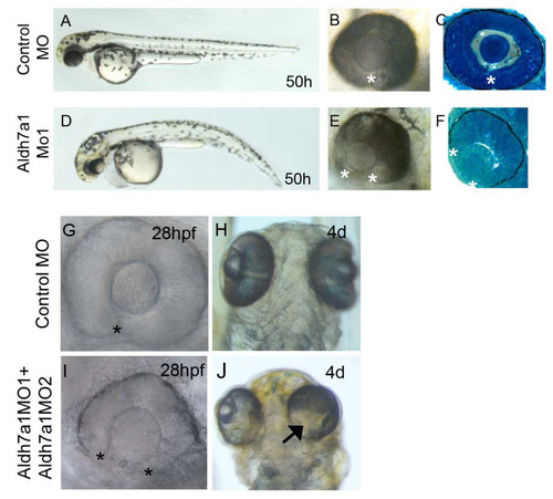
aldh7a1 loss-of-function phenotype. (A,D) Aldh7a1MO1 injected embryos developed bent tail compare to control MO injected embryos. (A-F) Lateral view of zebrafish eye at 50 hpf. Co-injection of Aldh7a1MO1 and Aldh7a1MO2 developed coloboma(I n=36/41,J n=25/31), compared to(G,H) control MO injected embryos.(C-F) Sagittal section of eye. Asterisks indicate edges of optic fissure, arrow indicates ventral defect in the eye at 4d. |
|
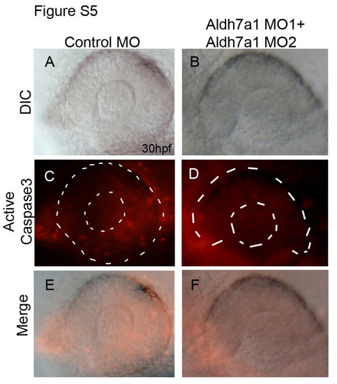
Coloboma in aldh7a1morphant fish is not due to apoptosis. Active Caspase3 staining of control (C,E) and aldh7a1 morphants embryos (D,F). White lines in C, D demarcates the eye. |

