- Title
-
Two additional midline barriers function with midline lefty1 expression to maintain asymmetric Nodal signaling during left-right axis specification in zebrafish
- Authors
- Lenhart, K.F., Lin, S.Y., Titus, T.A., Postlethwait, J.H., Burdine, R.D.
- Source
- Full text @ Development
|
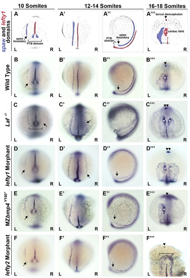
spaw and lefty1 expression phenotypes. (A-F′′)Time course of single color, double in situ hybridizations for spaw and lefty1 from 10-18 somites in posterior (A-F), mid-LPM (A′-F′), left lateral (A′-F′) and anterior LPM (A′′-F′′) views. (A-A′′) False-colored images of zebrafish embryos from B-B′′ depicting the domains of spaw and lefty1. In situ hybridizations of spaw or lefty1 alone confirm the reported phenotypes. Arrows, boundary of detectable spaw expression; brackets, ectopic spaw across the midline; arrowheads, lefty1 in the diencephalon; asterisks, lefty1 in the cardiac mesoderm. L, left; R, right. EXPRESSION / LABELING:
PHENOTYPE:
|
|
Characterization of the bmp4Y180* allele. (A) Bmp4 protein with location of the Y180* mutation indicated. RAKR, cleavage sequence. (B) Sequence comparisons of cDNA from wild type (wt) and bmp4Y180* mutants. (C-G) Zebrafish embryos at 24-36 hours post-fertilization (hpf). (C) WT embryo. (D) bmp4Y180* mutant with absent ventral fin (arrow) and cloaca defects (arrowhead). (E) Uninjected embryo. (F) Embryo injected with bmp4 RNA exhibiting V4 ventralization. (G) Embryo injected with bmp4Y180* RNA lacking morphological defects. PHENOTYPE:
|
|
Characterization of the novel bmp4S355* and bmp4C365S mutant alleles. (A) Diagram of the Bmp4 protein with locations of the S355* and C365S mutations indicated. (C,E) Sequence results from PCR amplification of the bmp4 gene from cDNA obtained from wild type (wt), bmp4C365S (C) and bmp4S355* (E) mutants confirming the identity and location of genetic lesions. (B,D,F-J) Brightfield images of zebrafish at 24-36 hpf. (B) WT embryo. (D) bmp4C365S mutant with no obvious defects in the ventral fin or cloaca. (F) bmp4S355* mutant exhibiting significant loss of the ventral fin (arrow). (G) Uninjected embryo. (H) Embryo injected with 50 pg of S355* RNA exhibiting no morphological defects. (I,J) Embryos injected with 50 pg of C365S RNA displaying C2-C3 (I) and C4 (J) dorsalization. Images of WT and uninjected embryos are duplicated from Fig. 2. PHENOTYPE:
|

Unillustrated author statements PHENOTYPE:
|


