- Title
-
Ret signalling integrates a craniofacial muscle module during development
- Authors
- Knight, R.D., Mebus, K., d'Angelo, A., Yokoya, K., Heanue, T., and Roehl, H.
- Source
- Full text @ Development
|
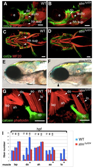
Musculoskeletal labelling of stmhy024 zebrafish mutants reveals specific muscle defects. (A-D) Immunolabelling of muscle (red) and cartilage (green) reveals that stmhy024 mutants (B,D) have reduced dilator operculi (do), adductor operculi (ao), levator operculi (lo) and hyohyoideus superiores (hh sup) muscles (arrowheads) relative to wild type (WT; A,C). e, eye. (E,F) stmhy024 mutants (F) have tightly closed mouths (arrow) relative to WT (E). (G,H) Higher magnification views of dorsal pharyngeal arch muscles reveal that the do, ao and lo attaching to the opercular bone (op, green) are reduced in stmhy024; the non-opercular muscles levator arcus palatini (lap) and adductor hyoideous (ah) are unaffected. (I) Quantification of fibre number in dorsal arch muscles (mean ± s.e.m.) reveals a specific and significant reduction (*P<0.0001) of the opercular muscles in stmhy024 relative to WT (n=10), during muscle development at 72, 96 and 120 hours post-fertilisation (hpf). Scale bars: 100 μm in A-D; 50 μm in G,H. |
|
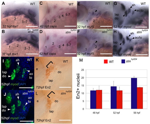
stmhy024 mutants show myogenic defects specifically in opercular muscles. (A-H) In situ hybridisation with tbx1 (A,B) and capsulin (C,D) probes reveals that muscle specification is unaffected in the pharyngeal arch muscle precursors of stmhy024. However, both myf5 (E,F) and myod (G,H) expression are reduced in stmhy024 relative to wild type (WT). Arrowheads indicate muscle primordia. (I-L) Fluorescent in situ reveals that myod expression is reduced specifically in the precursors of the lo and ao at 52 hours post-fertilisation (hpf; I,J) but is unaffected in the adjacent ah. At later stages, there are fewer En2-expressing (En2+) muscle cells specifically in the do but not the lap of stmhy024 (K,L). Brackets indicate do muscle cell nuclei. (M) Quantification of En2+ muscle precursors in stmhy024 (blue) and WT (red) at 48, 52 and 56 hpf reveals that stmhy024 mutants have fewer muscle precursor cells than WT in the lap or do primordia from 56 hpf (mean ± s.e.m.). Pharyngeal arches 1-7 are shown by numbering in A-F. ah, adductor hyoideous; ao, adductor operculi; do, dilator operculi; e, eye; lap, levator arcus palatini; lo, levator operculi. Scale bars: 50 μm in A-H,K-L; 20 μm in I,J. EXPRESSION / LABELING:
PHENOTYPE:
|
|
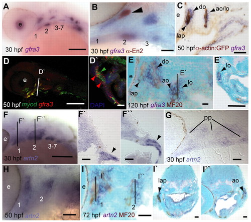
gfra3 and artn2 show tissue-specific expression in an opercular complex. (A,B) gfra3 (purple) is expressed in pharyngeal mesoderm at 30 hours post-fertilisation (hpf; A) and in muscle precursors (arrowhead) with En2 (brown, B). (C) At 50 hpf, gfra3 is expressed in forming muscle fibres expressing GFP (brown) in alpha-actin:GFP animals and associated cells. (D,D′) Fluorescent in situ of myod (green) and gfra3 (red) reveals colocalisation (yellow) in all forming pharyngeal arch muscles, but not in fin or ocular muscles. A transverse section (indicated by the line in D) through the second arch reveals gfra3 expression in myod- cells (D′). Red and green arrowheads indicate gfra3+myod? and gfra3+myod+ cells, respectively. (E,E′) At stages when primary muscle fibres have formed (120 hpf), sagittal sections show that gfra3 is expressed in mesenchymal cells adjacent to muscle fibres (brown, E) that completely surround the forming lo muscle (E′). Line in E indicates level of section in E′. (F-I′′) In situ hybridisation shows that artn2 is expressed in mesenchyme of the pharyngeal arches from 30 hpf. Transverse (F′,F′′) and sagittal (G) sections of the arches (1,2), reveal that artn2 expression is restricted to mesenchymal cells (arrowheads) and is excluded from the pharyngeal pouches (pp). Subsequently, artn2 becomes restricted to discrete domains in the dorsal first and second arches at 50 hpf (H). Sagittal (I) and transverse (I′,I*) sections reveal that as muscle fibres form (brown), artn2 expression is adjacent to the forming opercular muscles. Lines in I indicate level of sections in I′ and I*. Pharyngeal arches 1-7 are shown by numbering. ao, adductor operculi; do, dilator operculi; e, eye; lap, levator arcus palatini; lo, levator operculi. Scale bars: 100 μm in A,D,E′,F,I′,I* 20 μm in B,C,D′,E,F′-I. |
|
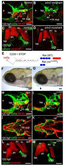
Loss of Ret or Artn2 function results in specific opercular muscle defects. (A-D,H-M) Immunolabelling of cartilage, bone (green) and muscles (red) of uninjected controls (A,C), artn2 morphants (B,D), wild type (WT; H,J,L) and rethu2846 mutants (I,K,M) at 120 hours post-fertilisation (hpf). Specific reductions of the do, ao, lo and hh sup muscles occur in artn2 morphants and rethu2846 mutants, but the lap and ah muscles are unaffected. (E-G) rethu2846 mutants have a tightly closed mouth (G, arrowhead) relative to WT (F) and have a point mutation in codon 229 (T>A) of exon 4 in the ret gene that changes a cysteine to a stop codon in the coding sequence (E). This results in a truncated protein lacking the tyrosine kinase domain (red), the transmembrane region (green) and part of the extracellular domain (blue). A morpholino directed to the exon 1 splice donor site (retSp) causes aberrant splicing of ret. ah, adductor hyoideous; ao, adductor operculi; do, dilator operculi; e, eye; hh sup, hyohyoideus superiores; lap, levator arcus palatini; lo, levator operculi. Scale bars: 100 μm in A,B,H-K; 50 μm in C,D,L,M. PHENOTYPE:
|
|
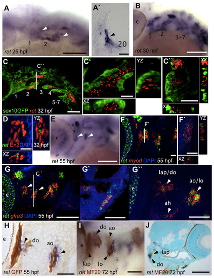
ret expression becomes restricted to cells associated with opercular muscles. (A-B) ret is expressed in the first (1) and second (2) arches at 25 hours post-fertilisation (hpf; A, white arrowheads); transverse section through the arch (A′) reveals that ret+ cells (black arrowhead) lie medially. ret becomes restricted to medial cells of the arches at 30 hpf (B) relative to GFP+ cells in sox10:GFP animals (C). (C-D) Confocal sections reveal that ret+ cells are surrounded by GFP+ cells (C′) and transverse sections fail to show colocalisation of ret and GFP (C′′). At similar stages, ret expression is detected in myoblasts of the first arch that show En2 immunoreactivity (D). (E) ret expression in the arches becomes restricted to discrete populations of cells, including some associated with the developing opercular muscles (arrowheads). (F,F′) ret and myod do not appear to be expressed in the same cells of the opercular muscles (F) and transverse sections fail to show colocalisation (F′). (G-G′′) ret and gfra3 are both expressed in opercular muscles at 55 hpf (G) and transverse sections (G′) and ventral views (G′′) show that they colocalise in the ao and lo, but not in the ah. (H) Differentiating muscle fibres (brown) in alpha-actin:GFP fish do not express ret (blue) at 55 hpf, but ret+ cells lie adjacent to fibres (arrowheads). (I,J) Lateral view of the arches in 72 hpf embryos following in situ hybridisation and immunolabelling (I) shows that ret expressing cells (blue) are associated with the forming do, ao and lo muscles (brown). Transverse sections reveal that ret is expressed in cells of the do but not the lap (J) at this stage. Lines in C, F and G indicate the level of section in C′′, F′ and G′, respectively. yz and xz planes are shown for C′, C′′, D and F′. ah, adductor hyoideous; ao, adductor operculi; do, dilator operculi; e, eye; lap, levator arcus palatini; lo, levator operculi. Pharyngeal arches 1-7 are shown by numbering in A-C. Scale bars: 100 μm in A-C,E,G,J; 20 μm in A′,C′,C′′,F,F′,G′-I; 10 μm in D. EXPRESSION / LABELING:
|
|
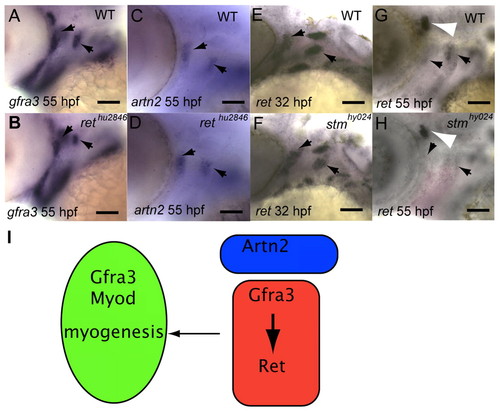
Gfra3 function is required for maintaining ret expression in opercular associated cells. (A-H) Expression of gfra3 (A,B), artn2 (C,D) and ret (E-H) in rethu2846 mutants (B,D) and wild-type (WT) siblings (A,C), stmhy024 mutants (F,H) and WT siblings (E,G) at 32 hours post-fertilisation (hpf; E,F) or 55 hpf (A-D,G,H). At 55 hpf, there are fewer gfra3-expressing cells in the forming adductor operculi (ao) and levator operculi (lo) (arrowheads) of rethu2846 mutants (B) compared with WT (A). By contrast, artn2 expression in the dorsal pharyngeal arches is unaffected in rethu2846 mutants (D) compared with WT (C). At 32 hpf, there are fewer ret+ cells (black arrowheads) in the dorsal second arch of stmhy024 mutants (F) relative to WT (E). Later, at 55 hpf ret expression in the opercular muscles is lost, but is unaffected in the anterior lateral line ganglia (white arrowhead, H). (I) A model for Ret signalling during opercular muscle development. Ret receptor and the Gfra3 co-receptor are present on myoblast cells (red). Artn2 from adjacent mesenchymal cells (blue) binds the Gfra3 co-receptor in Ret+ cells and muscle cells. Activation of Ret signalling by Gfra3 and Artn2 is required to maintain ret expression in myoblast cells and for myogenic differentiation to occur, involving myod expression and a loss of ret expression. Scale bars: 100 μm. |
|
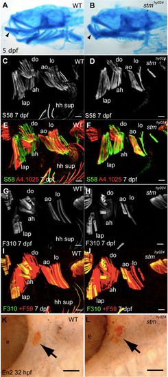
stmhy024 mutants have tightly closed mouths and do not show alterations to muscle identity. (A,B) Alcian Blue stained cranial cartilages show a normal morphology in stmhy024 mutants (B), but the mouth is tightly closed, changing the angle of the lower jaw (arrowhead) relative to wild type (WT; A). (C-J) The distribution of slow myosin (S58, C-F) fibres in the do, ao and lo is unchanged in stmhy024 (D,F) compared with WT (C,E) at 7 dpf, despite the reduction in fibre number (co-labelled by A4.1025; E,F). Likewise, fast myosin (F310, G-J) fibres show a similar distribution in dorsal arch muscles of stmhy024 (H,J) relative to WT (G,I) at 7 dpf. A comparison of F310 to all fibres (+F59, I,J) reveals that the reduced number of muscle fibres in opercular muscles stmhy024 does not involve a specific loss of fast fibres (I,J). (K,L) Engrailed 2 protein (En2) expression (arrows) occurs in the lap/do primordia at 32 hpf as normal in stmhy024 (K) and WT siblings (L). ah, adductor hyoideous; ao, adductor operculi; do, dilator operculi; e, eye; hh sup, hyohyoideus superiores; lap, levator arcus palatini; lo, levator operculi. Scale bars: 20 μm in C-J; 50 μm in K,L. |
|
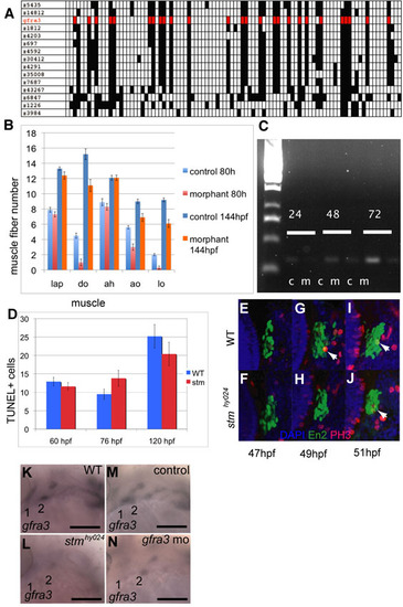
Gfra3 function is required for opercular muscle development. (A) Radiation hybrid panel mapping of zebrafish gfra3 (red) reveals that it segregates with other genes lying between z1812 and z5435 in agreement with SSLP mapping data. Filled boxes represent a positive result from the PCR on pooled hybridoma cells containing defined regions of the zebrafish genome. (B) Quantification of muscle fibers in gfra3 morphants reveals a statistically significant reduction of opercular muscles similar to stmhy024 mutants (n=10) but also reveals that muscles recover between 80 hpf to 144 hpf in gfra3 morphants. Error bars represent s.e.m. (C) RT-PCR amplification of a 380 bp fragment of gfra3 using primers designed to exons 2 and 4 from animals injected by gfra3 morpholinos (m) reveals a reduction of spliced gfra3 RNA compared to uninjected controls (c) at 24, 48 and 72 hpf. (D) TUNEL labelling of cells undergoing programmed cell death in the pharyngeal arches revealed no significant differences (P<0.01) between stmhy024 and wild type (WT) at 60, 76 and 120 hpf. Error bars represent s.e.m. (E-J) Very few muscle precursors of the lap and do expressing En2 (green) are labelled by anti-phosphohistone3 (PH3, red), a marker of proliferating cells in WT (E,G,I) or stmhy024 mutants (F,H,J) at 47, 49 and 51 hpf relative to DAPI (blue). There appears to be no change to the number of proliferating En2+ stained nuclei (arrowheads) in stmhy024 mutants, although there are fewer En2+ cells overall by 51 hpf. (K-N) gfra3 expression is reduced in the arches (1,2) of stmhy024 (L) at 25 hpf relative to WT (K) and, likewise, is reduced in gfra3 morphants (N) at 25 hpf relative to uninjected control animals (M). |

ret-expressing cells in the opercular muscles are not derived from neural crest (NC) and connective cell markers are present in Ret signalling mutants. (A,B) Injection of morpholinos to tfap2a and tfap2c results in a loss of NC cells (data not shown), but ret is still expressed in the arches (1,2) of tfap2+c morphants (B) compared with wild type (WT; A). (C,C′) A comparison of ret expression in the arches (1,2) to GFP+ NC cells in fli1:GFP animals (C), shows no expression of ret in NC cells. In the second arch the ret+ cells are surrounded by GFP+ cells, which suggests that they are mesodermal (C′). (D,E) Connective tissues and opercular bone (op, arrowhead) labelled by zns-5 appear normal in rethu2846 mutants (D) compared with WT (E). (F,G) ten-w expression in presumptive connective tissues (arrowhead) associated with the forming opercular bone (op) is unchanged in stmhy024 mutants (F) at 65 hpf compared with WT (G). (H,I) At 4 dpf, ten-w-expressing cells on the opercular bone are associated with the ao and lo muscles (arrowheads) and are likely to be tendon precursors. Expression of ten-w is still present in these tendon cells in rethu2846 mutants (I) relative to WT (H), but is reduced, reflecting the reduced number of muscle fibres connecting to the bone. (J-M) At 3 dpf and onwards, ten-c expression is in presumptive connective cells (arrowheads) associated with the opercular bone. ten-c expression in these cells is not obviously reduced in either stmhy024 mutants (K) or rethu2846 mutants (M) compared with WT (J,L). ah, adductor hyoideous; allg, anterior lateral line ganglia; ao, adductor operculi; e, eye; lo, levator operculi. Scale bars: 100 μm in A-E; 20 μm in C′,F-M. |

