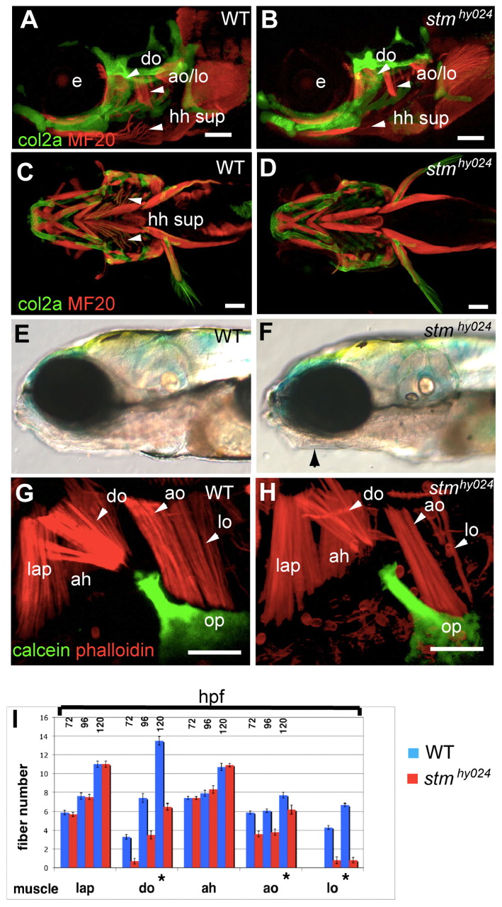
Fig. 1 Musculoskeletal labelling of stmhy024 zebrafish mutants reveals specific muscle defects. (A-D) Immunolabelling of muscle (red) and cartilage (green) reveals that stmhy024 mutants (B,D) have reduced dilator operculi (do), adductor operculi (ao), levator operculi (lo) and hyohyoideus superiores (hh sup) muscles (arrowheads) relative to wild type (WT; A,C). e, eye. (E,F) stmhy024 mutants (F) have tightly closed mouths (arrow) relative to WT (E). (G,H) Higher magnification views of dorsal pharyngeal arch muscles reveal that the do, ao and lo attaching to the opercular bone (op, green) are reduced in stmhy024; the non-opercular muscles levator arcus palatini (lap) and adductor hyoideous (ah) are unaffected. (I) Quantification of fibre number in dorsal arch muscles (mean ± s.e.m.) reveals a specific and significant reduction (*P<0.0001) of the opercular muscles in stmhy024 relative to WT (n=10), during muscle development at 72, 96 and 120 hours post-fertilisation (hpf). Scale bars: 100 μm in A-D; 50 μm in G,H.
Image
Figure Caption
Figure Data
Acknowledgments
This image is the copyrighted work of the attributed author or publisher, and
ZFIN has permission only to display this image to its users.
Additional permissions should be obtained from the applicable author or publisher of the image.
Full text @ Development

