Fig. S1
|
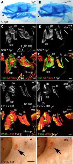
stmhy024 mutants have tightly closed mouths and do not show alterations to muscle identity. (A,B) Alcian Blue stained cranial cartilages show a normal morphology in stmhy024 mutants (B), but the mouth is tightly closed, changing the angle of the lower jaw (arrowhead) relative to wild type (WT; A). (C-J) The distribution of slow myosin (S58, C-F) fibres in the do, ao and lo is unchanged in stmhy024 (D,F) compared with WT (C,E) at 7 dpf, despite the reduction in fibre number (co-labelled by A4.1025; E,F). Likewise, fast myosin (F310, G-J) fibres show a similar distribution in dorsal arch muscles of stmhy024 (H,J) relative to WT (G,I) at 7 dpf. A comparison of F310 to all fibres (+F59, I,J) reveals that the reduced number of muscle fibres in opercular muscles stmhy024 does not involve a specific loss of fast fibres (I,J). (K,L) Engrailed 2 protein (En2) expression (arrows) occurs in the lap/do primordia at 32 hpf as normal in stmhy024 (K) and WT siblings (L). ah, adductor hyoideous; ao, adductor operculi; do, dilator operculi; e, eye; hh sup, hyohyoideus superiores; lap, levator arcus palatini; lo, levator operculi. Scale bars: 20 μm in C-J; 50 μm in K,L. |

