- Title
-
Reiterative roles for FGF signaling in the establishment of size and proportion of the zebrafish heart
- Authors
- Marques, S.R., Lee, Y., Poss, K.D., and Yelon, D.
- Source
- Full text @ Dev. Biol.
|
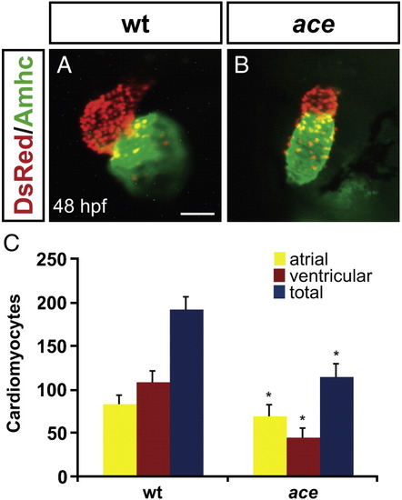
ace mutant embryos have reduced numbers of both ventricular and atrial cardiomyocytes. (A, B) Frontal views of hearts from wild-type (A) and ace mutant (B) embryos at 48 hpf; immunofluorescence detects DsRed (red) in all cardiomyocyte nuclei and atrial myosin heavy chain (Amhc; green) in atrial cells. (A) In a wild-type heart, the ventricle (red) and atrium (green) exhibit typical looping and expansion. (B) In an ace mutant heart, the chambers are unlooped and small, with a particularly apparent reduction of the ventricle. Scale bar represents 50 μm; both images are shown at the same magnification. (C) Quantification of cardiomyocytes at 48 hpf reveals that the numbers of both ventricular and atrial cells are significantly decreased in ace mutants, with ventricular cell number being more affected then atrial cell number. Graph indicates mean and standard deviation for each data set; asterisks indicate statistically significant differences relative to wild-type (p < 0.005, Student's t-test). n = 13 for wild-type, and n = 19 for ace mutants; see also Supplemental Table 1. |
|
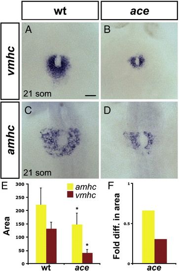
Chamber disproportionality is evident prior to heart tube assembly in ace mutants. (A?D) In situ hybridization depicts expression of vmhc (A, B) and amhc (C, D) at the 21-somite stage; dorsal views, anterior to the top. Scale bar represents 50 μm; all images are shown at the same magnification. (A) In wild-type embryos, vmhc is expressed in a ring of ventricular cardiomyocytes just prior to heart tube extension. (B) In ace mutant embryos, the population of vmhc-expressing cells is clearly reduced (n = 14/15). (C) amhc is expressed in a ring of atrial cardiomyocytes, surrounding the ventricular cardiomyocytes. (D) The population of amhc-expressing cells is also reduced in ace mutant embryos (n = 11/13). (E) Graph indicates mean and standard deviation of areas of expression (in μm2) of amhc and vmhc in wild-type and ace mutant embryos. Asterisks indicate statistically significant differences relative to wild-type (p < 0.005, Student's t-test). n e 10 for all data sets; see also Supplemental Table 2. (F) Graph indicates fold difference in mean areas of gene expression relative to wild-type. EXPRESSION / LABELING:
PHENOTYPE:
|
|
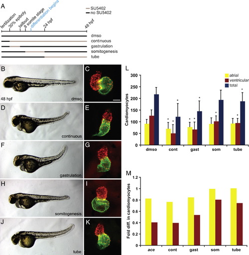
Transient reduction of FGF signaling leads to differential reduction of cardiomyocyte numbers. (A) Schematic depicts the transient periods of FGF signaling inhibition caused by addition and removal of SU5402. Black lines represent time intervals with normal FGF signaling, and tan lines represent intervals of SU5402 treatment. Control embryos were treated only with DMSO, ?continuous? SU5402 treatment extended from 30% epiboly (3 hpf) until 48 hpf, ?gastrulation? treatment began at 30% epiboly and ended at the tailbud stage (10 hpf), ?somitogenesis? treatment began at the 8-somite stage (13 hpf) and ended at 24 hpf, and ?tube? treatment extended from 24 hpf to 48 hpf. (B?K) Representative 48 hpf embryos, lateral views, exhibiting morphology resulting from each type of SU5402 treatment, coupled with respective frontal views of hearts in which immunofluorescence detects DsRed (red) in all cardiomyocyte nuclei and Amhc (green) in atrial cells, as in Fig. 1. Scale bar represents 50 μm; all images of hearts are shown at the same magnification. Embryos treated continuously or during gastrulation occasionally exhibited severe tail truncations characteristic of higher SU5402 concentrations (Griffin and Kimelman, 2003); severely truncated embryos were excluded from quantification of cardiomyocytes. The concentration of SU5402 used did not induce nonspecific apoptosis, as indicated by TUNEL (SRM and DY, unpublished results). (L) Quantification of cardiomyocytes at 48 hpf after each type of SU5402 treatment. Graph indicates mean and standard deviation for each data set; asterisks indicate statistically significant differences relative to DMSO-treated controls (p < 0.005, Student's t-test). Note that the total number of cardiomyocytes following somitogenesis treatment is also significantly different from the control number, although with a larger p value (p < 0.01). (M) Graph indicates fold difference in mean values relative to wild-type for ace mutant embryos and embryos treated with SU5402. n = 33 for DMSO, n = 8 for continuous treatment, n = 28 for gastrulation treatment, n = 28 for somitogenesis treatment, and n = 22 for tube treatment; see also Supplemental Table 3. EXPRESSION / LABELING:
|
|
Heat shock of Tg(hsp70:ca-fgfr1) embryos causes ectopic FGF signaling and perturbs pectoral fin development. (A, B) In situ hybridization depicts pea3 expression at 32 hpf; dorsal views, anterior to the left. (A) After heat shock at 24 hpf, non-transgenic (non-tg) embryos show a wild-type restriction of pea3 expression to specific regions, including the midbrain?hindbrain boundary and the pharyngeal pouches. (B) In contrast, heat shock of transgenic (tg) embryos causes strong and ectopic expression of pea3. (C, D) Dorsal views at 72 hpf of embryos after heat shock at the 8-somite stage. (C) Non-transgenic embryos exhibit pectoral fins of normal length (bar). (D) Transgenic embryos lack at least one pectoral fin, as shown here (n = 14/50), or have two small fins (n = 31/50). EXPRESSION / LABELING:
|
|
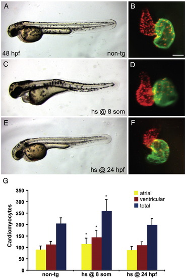
Increased FGF signaling prior to terminal differentiation induces a surplus of cardiomyocytes. (A?F) Representative non-transgenic (A) or Tg(hsp70:ca-fgfr1) (C, E) sibling embryos at 48 hpf, lateral views, exhibiting morphology resulting from heat shock at the 8-somite stage (C) or at 24 hpf (E), coupled with respective frontal views of hearts in which immunofluorescence detects DsRed (red) in all cardiomyocyte nuclei and Amhc (green) in atrial cells, as in Fig. 1. Scale bar represents 50 μm; all images of hearts are shown at the same magnification. (C, D) Although heat shock at the 8-somite stage causes pericardial edema (C) and dysmorphic cardiac chambers (D), heat shock at 24 hpf does neither (E, F). (G) Quantification of cardiomyocytes at 48 hpf reveals that increased FGF signaling causes a significant cardiomyocyte surplus only in embryos heat shocked at the 8-somite stage. Graph indicates mean and standard deviation for each data set; asterisks indicate statistically significant differences relative to non-tg controls (p < 0.005, Student's t-test). n = 24 for non-tg, n = 18 for heat shock at the 8-somite stage, and n = 14 for heat shock at 24 hpf; see also Supplemental Table 5. EXPRESSION / LABELING:
|
|
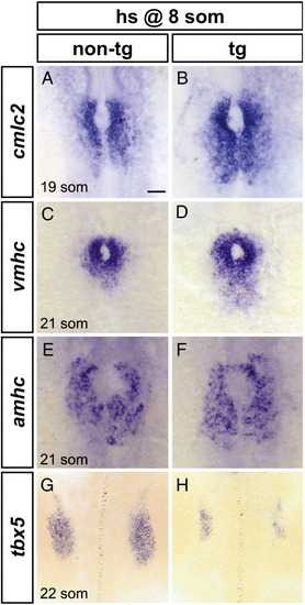
Increased FGF signaling enhances formation of cardiomyocyte populations and inhibits formation of the pectoral fin field. (A?H) In situ hybridization depicts expression of cmlc2, vmhc, amhc and tbx5 at the 19-somite or 21-somite stage in non-transgenic (A, C, E, G) and Tg(hsp70:ca-fgfr1) (B, D, F, H) sibling embryos after heat shock at the 8-somite stage; dorsal views, anterior to the top. Scale bar represents 50 μm; all images are shown at the same magnification. (A, B) Ectopic FGF signaling leads to a posterior expansion of cmlc2-expressing cardiomyocytes (n = 30/37). (C, D) Ectopic FGF signaling leads to a posterior expansion of vmhc-expressing ventricular cardiomyocytes (n = 16/19). (E, F) Ectopic FGF signaling leads to a mild posterior expansion of amhc-expressing atrial cardiomyocytes (n = 8/10). (G, H) Ectopic FGF signaling leads to reduction of the pectoral fin field, as demarcated by tbx5 expression (n = 18/18). EXPRESSION / LABELING:
|
|
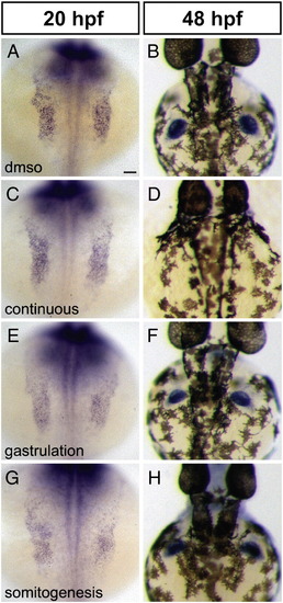
Reduction of FGF signaling does not expand the forelimb field. (A?H) In situ hybridization depicts expression of tbx5 at 20 hpf (A, C, E, G) and 48 hpf (B, D, F, H); dorsal views, anterior to the top. Scale bar represents 50 μm; all images are shown at the same magnification. (A?D) Compared to control embryos treated only with DMSO (A, B), embryos exposed to continuous SU5402 treatment exhibit normal areas of tbx5 expression in the pectoral fin fields at 20 hpf (C; n = 8/8) but lack pectoral fins at 48 hpf (D; n = 15/15). (E, F) Embryos treated with SU5402 during gastrulation exhibit normal areas of tbx5 expression in the pectoral fin fields at 20 hpf (n = 16/16) and normal pectoral fins at 48 hpf (n = 18/18). (G, H) Embryos treated with SU5402 during somitogenesis do not display increased areas of tbx5 expression in the pectoral fin fields at 20 hpf (n = 9/9) and do not have detectable defects in pectoral fin size at 48 hpf (n = 15/16), although fin morphology varies between treated embryos. It is noteworthy that the level of tbx5 expression generally appears higher in wild-type embryos than in SU5402-treated embryos. EXPRESSION / LABELING:
|
Reprinted from Developmental Biology, 321(2), Marques, S.R., Lee, Y., Poss, K.D., and Yelon, D., Reiterative roles for FGF signaling in the establishment of size and proportion of the zebrafish heart, 397-406, Copyright (2008) with permission from Elsevier. Full text @ Dev. Biol.

