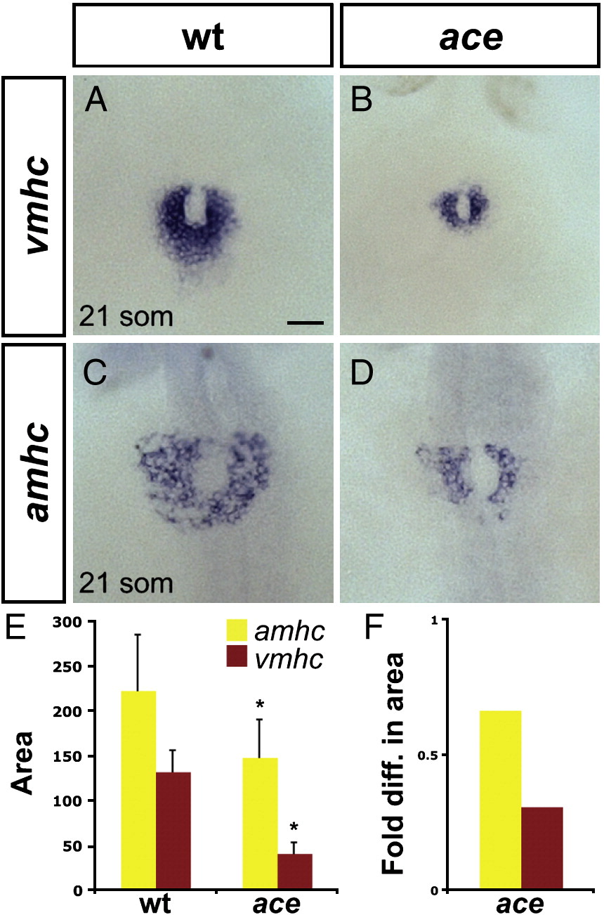
Fig. 2 Chamber disproportionality is evident prior to heart tube assembly in ace mutants. (A?D) In situ hybridization depicts expression of vmhc (A, B) and amhc (C, D) at the 21-somite stage; dorsal views, anterior to the top. Scale bar represents 50 μm; all images are shown at the same magnification. (A) In wild-type embryos, vmhc is expressed in a ring of ventricular cardiomyocytes just prior to heart tube extension. (B) In ace mutant embryos, the population of vmhc-expressing cells is clearly reduced (n = 14/15). (C) amhc is expressed in a ring of atrial cardiomyocytes, surrounding the ventricular cardiomyocytes. (D) The population of amhc-expressing cells is also reduced in ace mutant embryos (n = 11/13). (E) Graph indicates mean and standard deviation of areas of expression (in μm2) of amhc and vmhc in wild-type and ace mutant embryos. Asterisks indicate statistically significant differences relative to wild-type (p < 0.005, Student's t-test). n e 10 for all data sets; see also Supplemental Table 2. (F) Graph indicates fold difference in mean areas of gene expression relative to wild-type.
Reprinted from Developmental Biology, 321(2), Marques, S.R., Lee, Y., Poss, K.D., and Yelon, D., Reiterative roles for FGF signaling in the establishment of size and proportion of the zebrafish heart, 397-406, Copyright (2008) with permission from Elsevier. Full text @ Dev. Biol.

