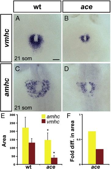
Chamber disproportionality is evident prior to heart tube assembly in ace mutants. (A?D) In situ hybridization depicts expression of vmhc (A, B) and amhc (C, D) at the 21-somite stage; dorsal views, anterior to the top. Scale bar represents 50 μm; all images are shown at the same magnification. (A) In wild-type embryos, vmhc is expressed in a ring of ventricular cardiomyocytes just prior to heart tube extension. (B) In ace mutant embryos, the population of vmhc-expressing cells is clearly reduced (n = 14/15). (C) amhc is expressed in a ring of atrial cardiomyocytes, surrounding the ventricular cardiomyocytes. (D) The population of amhc-expressing cells is also reduced in ace mutant embryos (n = 11/13). (E) Graph indicates mean and standard deviation of areas of expression (in μm2) of amhc and vmhc in wild-type and ace mutant embryos. Asterisks indicate statistically significant differences relative to wild-type (p < 0.005, Student's t-test). n e 10 for all data sets; see also Supplemental Table 2. (F) Graph indicates fold difference in mean areas of gene expression relative to wild-type.
|
