- Title
-
Hereditary spherocytosis in zebrafish riesling illustrates evolution of erythroid ß-spectrin structure, and function in red cell morphogenesis and membrane stability
- Authors
- Liao, E.C., Paw, B.H., Peters, L.L., Zapata, A., Pratt, S.J., Do, C.P., Lieschke, G., and Zon, L.I.
- Source
- Full text @ Development
|
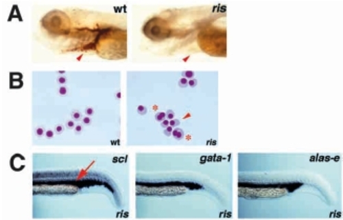
Embryonic blood defects in ris. In this and all subsequent figures embryos are shown as lateral views, with anterior left and ventral down, at 10X magnification, unless specified otherwise. (A) Whole-mount o-dianisidine staining of wildtype and ris embryos at 4 dpf, showing absence of blood cells in ris (arrowhead). (B) Wright- Giemsa staining of embryonic blood cells collected from wild-type and ris embryos at 2.5 dpf. Wild-type primitive erythrocytes are spherical and nucleated, whereas ris primitive erythrocytes are often abnormally shaped (arrowhead) and binucleated (asterisk). 20X magnification. (C) In situ analysis of ris embryos at 23 hours post-fertilization (hpf), with antisense riboprobes of scl, gata-1 and alas-e. At 23 hpf, blood cells are restricted to the intermediate cells mass (ICM), dorsal to the yolk tube extension (arrow). EXPRESSION / LABELING:
|
|
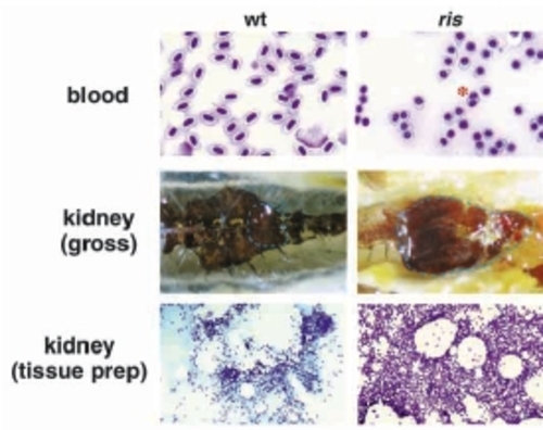
Adult ris blood and marrow. Peripheral blood and kidney collected from wild-type and ris adults. Blood smear and kidney tissue preparation were Wright-Giemsa stained, and whole kidneys are shown in situ, situated in the retroperitoneal cavity of the adult animal (organ margin demarcated with broken lines). Mutant erythrocytes are spherical or tear-drop shaped, with round nuclei. More bi-nucleated cells are seen in the mutant (asterisk). In contrast, the wild-type red cells and nuclei are elliptical. Mutant kidney tissue is dramatically enlarged compared with wild type, with increased cellularity. Blood, gross kidney and tissue kidney are shown at 20x, 4x and 4x, respectively. PHENOTYPE:
|
|
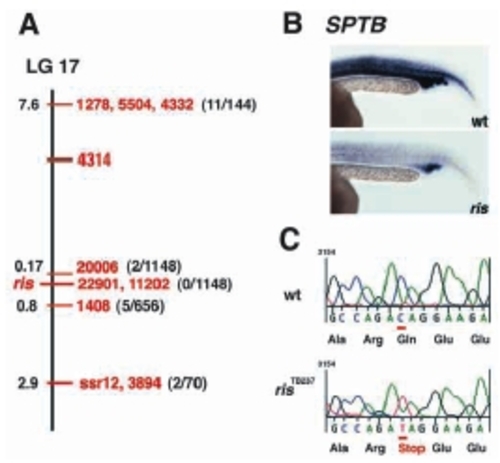
sptb is the defective gene in ris. (A) Genetic map of LG 17, showing a sample number of SSLP markers tested in the meiotic mapping (in red), and their relative distances from ris locus (in black). The number of recombinants/the total number of animals tested are indicated in parenthesis. (B) sptb whole-mount RNA in situ on wild-type and ris embryos at 23 hpf. Identical staining of ICM, myotube and somites is observed with riboprobes generated from BScd, BF1/238 (shown here), or BSPH clones. (C) Sequence of a five codon interval encompassing the C to T transversion mutation at position +3130 that was found in ris. EXPRESSION / LABELING:
|
|
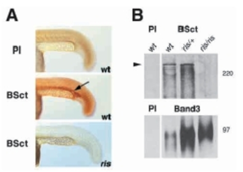
Sptb product is not detected in ris blood cells. (A) Wholemount immunohistochemical detection of Sptb product with anti- BSct polyclonal antisera in wild-type and ris embryos at 23 hpf. (B) Western blot detection of Sptb from whole-blood lysate collected from wild-type, ris/+ and ris/ris adult fish with anti-BSct polyclonal antisera (arrow), and normalized for sample loading with anti-Band3 polyclonal antisera. The zebrafish Sptb protein is >220 kDa in size. |
|
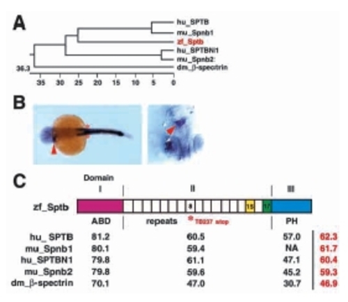
Phylogeny and sequence comparison of Sptb. (A) Phylogenetic tree of β-spectrin family members from Drosophila melanogaster, zebrafish, mouse and human. Numerical value indicates phylogenetic distance (= sum (residue distance) + (gaps x gaps penalty) + (gap residue x gap length penalty)). Zebrafish sptb is shown in red. (B) Left panel shows dorsal view (4X) of a 23 hpf embryo, with sptb expression in the myotube (arrowhead) and somites (asterisk). Identical staining of ICM, myotube and somites is observed with riboprobes generated from BScd, BF1/238 or BSPH (shown here) clones. Right panel shows ventral view of the myotube (arrowhead), with head pointing down. (C) Diagrammatic depiction of Sptb. Percentage identity between domains of β-spectrin homologs are indicated in black, where overall percentage identity is indicated in red. Domain II consists of 17 spectrin repeats arranged in coiled-coil helices. Repeat 15 (yellow shade) is required for binding to ankyrin, which in turn binds to band 3 (anion exchanger AE1). Repeat 17 (green shade) is required for ?head to head? association of αβ spectrin dimers. Domain I encompasses the actin binding domain (ABD) and is responsible for interactions with actin, adducin, centractin (ARP1 dynein), microtubule and protein 4.1 (and thereby glycophorin C). Domain III consists of the PH domain, which interacts with phosphotidylinositol-(4,5)-bisphosphate, βϒ G proteins, and protein kinase C. A C to T transversion mutation resulting in premature translation termination in repeat 8 in sptb (asterisk) is the molecular defect of the ris TB237 allele. |
|
Cell membrane and microtubule marginal band defects of ris erythrocytes. SEM and TEM analysis of peripheral blood cells. Wild-type erythrocytes are elliptical with smooth biconcave membrane surface. A central nuclear bulge (arrow) located within the biconcavity of wildtype erythrocytes corresponds to the nucleus. Heterozygote ris/+ erythrocytes are elliptical, but the membrane surface appears irregular (asterisk) and biconcavity is lost (arrowheads). Homozygote ris/ris red cells are smaller, spherocytic, and show profound surface pitting and spiculated membrane projections (asterisk). TEM analysis at 3000x magnicication, shows that the MB is situated adjacent to the lipid bilayer (arrows). Wild-type or ris/+ (not shown) MB consists of 20-24 microtubules, whereas ris MB consists of only 12-14 microtubule filaments. |
|
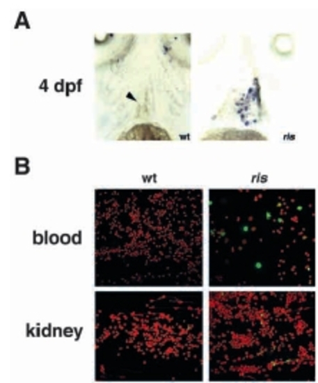
Apoptosis in ris embryonic and adult erythrocytes. (A) Colormetric TUNEL assay of wild-type and ris embryos at 4 dpf, shown in ventral view with head up. 10x magnification. No staining is observed in cardiac chamber (arrowhead) of wild-type embryo, whereas marked apoptosis is observed in red cells pooled in cardiac chamber of ris. (B) Fluorescence confocal TUNEL analysis of kidney and peripheral blood from wild-type and ris adults. The number of erythrocytes is severely reduced in the ris peripheral blood, where a larger fraction is undergoing apoptosis (green cells). The kidney of ris is hyper-cellular, with low level of apoptosis (green). |

