
|
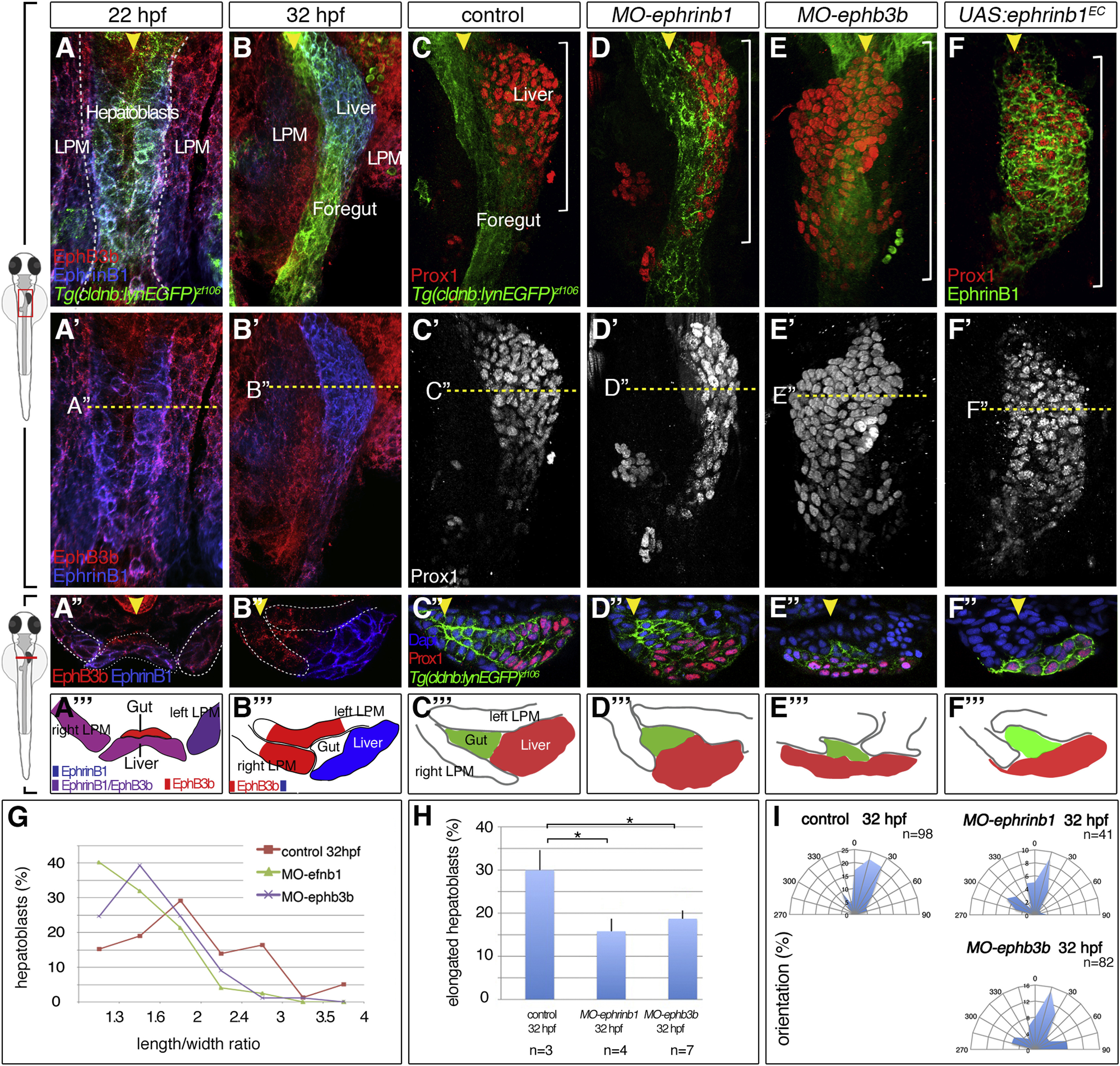
Fig. 4
Complementary EphrinB1 and EphB3b Expression Controls Hepatoblast Positioning and Cell-Shape Changes in Liver Bud Formation
(A-F?) At 22 hpf, EphrinB1 and EphB3b expression largely overlaps in future hepatoblasts and the LPM (A-A?). Complementary expression of both factors coincides with the start of liver budding: EphrinB1 in hepatoblasts and EphB3b in the gut and LPM (B?B?). At 32 hpf, hepatoblasts are located more posteriorly and medially compared with controls in MO-ephrinb1 (C-D?), in MO-ephb3b (E-E?), and upon conditional UAS:ephrinb1EC expression (F-F?). (A-F?) ventral views of confocal projections, anterior to the top; (A??F?) transverse sections of the foregut, as indicated by the dashed line in (A?-F?), and matching schematics (A?-F?); yellow arrowheads specify the midline and white brackets the length of the Prox1 domain.
(G?I) Cell shapes were determined with EphrinB1-staining at 32 hpf (see Figure 1F). Quantification of hepatoblast shape in control, MO-ephrinb1, and MO-ephb3b embryos: (G) L/W distribution for one representative bud; (H) proportion of elongated cells per bud; SEs are shown; and (I) orientation of elongated hepatoblasts with respect to the anteroposterior axis. ?p < 0.05.
See also Figure S2.
Reprinted from Developmental Cell, 39, Cayuso, J., Dzementsei, A., Fischer, J.C., Karemore, G., Caviglia, S., Bartholdson, J., Wright, G.J., Ober, E.A., EphrinB1/EphB3b Coordinate Bidirectional Epithelial-Mesenchymal Interactions Controlling Liver Morphogenesis and Laterality, 316-328, Copyright (2016) with permission from Elsevier. Full text @ Dev. Cell