
Fig. 9
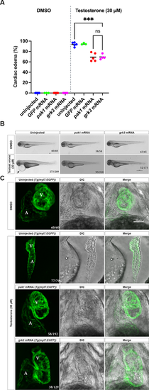
The testosterone phenotype is partially rescued by overexpression of pak1 or grk3 mRNA. (A) Wild-type embryos were injected with gfp, pak1 or grk3 mRNA. Embryos were exposed to vehicle or testosterone. Morphological phenotypes were assayed at 3 days post-fertilization. pak1 and grk3 mRNA reduced cardiac edema after testosterone exposure. Horizontal lines are the mean of each clutch. ****P<0.0001; ns, not significant (P>0.05); one-way ANOVA with Tukey?s multiple comparisons test. (B) Representative bright-field images of embryos from A at 3 days post-fertilization (dpf). Arrows indicate cardiac edema. Lateral views with anterior to the left, dorsal to the top. Fractions in the bottom right corners refer to the number of embryos with the indicated phenotype over the total number of embryos examined. Scale bar: 500 ?m. (C) Cardiac phenotypes examined on the background of Tg(myl7:EGFP) embryos after exposure to testosterone (30 �M) or vehicle controls (0.1% DMSO), beginning at 3 h post-fertilization. Representative confocal maximum intensity projections of live embryo hearts at 3 dpf. Fractions in the bottom right corners refer to the number of embryos with the indicated phenotype over the total number of embryos examined (30-75 embryos per clutch, two or three clutches per group). Myocardial cells are labeled in green; A, atrium; V, ventricle; DIC, differential interference contrast. Scale bar: 50 ?m. |