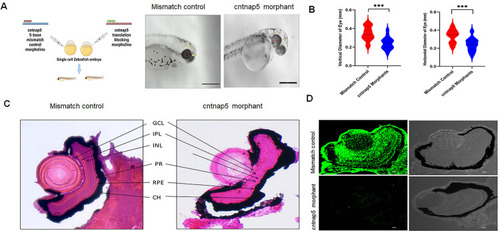
A. Morpholino knockdown of cntnap5a and 5b, left cartoon is created with BioRender.com licensed version. Zebrafish were microinjected with a cntnap5 translation blocking morpholinos. Images taken 96 hpf, eye of a mismatch morphant (left) and an eye cntnap5 morpholino injected zebrafish (right) showing smaller anatomical eye than mismatch control. B. Quantitative gross anatomical diameters along both the nasal-temporal axis (horizontal) and dorsal-ventral axis (vertical) of eyes measured from cntnap5 translation-blocking morpholino injected embryos and mismatch morpholino controls zebrafish at 96 hours post fertilization stage. bars = mean ± SD, ns not significant, ***p < 0.005. C. Histological analysis of zebrafish eye microinjected with mismatch morpholino (left) a cntnap5 translation blocking morpholino (right) using H&E stain at 96 hpf stage showing disrupted retinal layers in cntnap5 morphant. GCL, ganglion cell layer; IPL, inner plexiform layer; INL, inner nuclear layer; PR, photoreceptor layer; RPE, retinal pigment epithelium; CH indicates choroid. D. Representative IF image of tissues stained with anti-cntnap5 showing cntnap5 expression in mismatch morphant (upper left; lower Transmitted Light Differential Interference Contrast (TD) image) but no expression in a cntnap5 translation blocking morphants (lower left: right: TD image).
|
