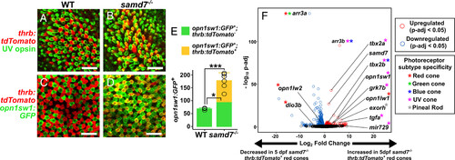
Red cones are transformed into hybrid red/UV cones in the larval samd7?/? retina. (A?D) Confocal images of 5-dpf WT and samd7?/? eyes show that UV opsin and UV-cone-specific opn1sw1:GFP is up-regulated in samd7?/? red cones (thrb:tdTomato+). (E) UV cones (opn1sw1:GFP+ cells per 1,600 �m2) are increased in the samd7?/? retina. The subset of UV cones that are not derived from red cones (opn1sw1:GFP+;thrb:tdTomato?) are also increased, suggesting that a small subset of supernumerary UV cones in the samd7?/? retina derive from another unknown cell type or precursor (mean � SD; n = 4 WT, n = 6 samd7?/?). (F) Volcano plot from bulk RNA-seq comparing gene expression in isolated 5-dpf WT thrb:tdTomato+ cells (i.e., red cones) and samd7?/? thrb:tdTomato+ cells (i.e., transformed hybrid red/UV cones). UV-cone genes are up-regulated in samd7?/? hybrid red/UV cones including tbx2a, tbx2b, opn1sw1, arr3b, mir729, tgfa, and grk7b (5). Red-cone-specific changes in gene expression were also observed, including reduced expression of the thyroid-hormone-inactivating enzyme, dio3b; increased expression of the red-shifted red opsin paralog, opn1lw1 (?max = 558 nm); and decreased expression of the blue-shifted red opsin paralog, opn1lw2 (?max = 548 nm). Similar changes in red opsin paralog expression were previously observed in larval zebrafish treated with thyroid hormone (44). Red/green-cone-specific arr3a expression was absent, and exorh and samd7 expression were increased in samd7?/? hybrid red/UV cones. (Scale bar, 10 �m.) Statistical comparisons in E were performed using the two-tailed, unpaired t test assuming unequal variance. *P < 0.05 and ***P < 0.0005. SD, standard deviation.
|
