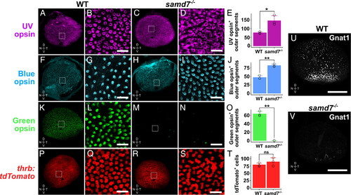
The numbers of blue and UV cones are approximately doubled, whereas green cones and rods are severely reduced in samd7?/? larvae. Confocal whole-mount images and high magnification insets of 5-dpf WT and samd7?/? eyes. (A?E) The number of UV cones stained with the UV opsin antibody is approximately doubled. (F?J) The number of blue cones stained with the blue opsin antibody is approximately doubled. (K?O) Green cones, as stained with the green opsin antibody, are absent from the samd7?/? eye. (P-T) The number of red cones, as identified with the thrb:tdTomato transgene, is unchanged, although the percentage of red cones that are directly contiguous appears to be increased in the mutant. (U and V) There is a severe reduction in the number of rods stained with the Gnat1 antibody, although a small population of rods with reduced Gnat1 signal can be observed in the ventral samd7?/? retina. (mean � SD; n = 3 per group). All fields of view quantified in (E, J, O, and T) were 40 � 40 �m2. Scale bars in all close-up views (B, D, G, I, L, N, Q, and S) = 10 �m. [Scale bar in (U and V), 100 �m.] D, V, N, T: Dorsal, Ventral, Nasal, Temporal. Statistical comparisons were performed using the two-tailed, unpaired t test assuming unequal variance. *P < 0.05, **P < 0.005. ns, not significant. SD, SD.
|
