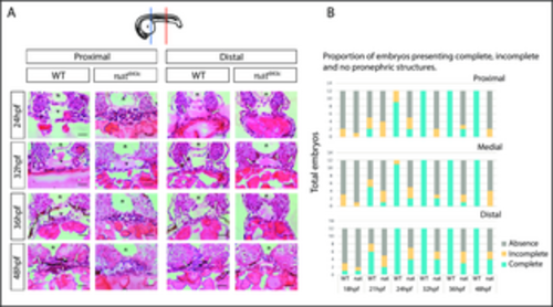
nattl43c mutants have a pronephric phenotype. A. Transversal H&E histological slides for Fn mutants and their WT siblings at proximal and distal levels. From 18 to 21 hpf no pronephric structures are visible. At 24, 32, 36, and 48 hpf, WT embryos pronephric structures (white arrows), as well as normal vascular structures (v) are noticeable. By 48 hpf WT embryos have normal intestinal lumen (i). Mutant embryos do not present normal vasculature or pronephric tubules, and by 48 hpf, mutants show poorly defined intestine and vasculature. Moreover, WT embryos show a great amount of melanocytic neural crest cells (arrowheads) whereas mutants have an exacerbated amount of blood cells. In mutants, the pronephric structure appears more frequently in distal levels than proximal levels, but they are poorly defined. n (notochord); v (vasculature); b (blood cells); i (intestine). Dorsal up. Scale bar: 15 ?m. n = 12 from three independent experiments (4 embryos each). The diagram shows the level examined: blue for proximal and red for distal. B. The proportion of embryos showing complete (bilateral appearance of evident and easily recognizable pronephric tubules), incomplete (absence of one structure), or total absence (no discernible or absence) of pronephric structures in three distinctive anatomical locations (proximal, medial, distal) as examined in H&E histology. n = 12.
|
