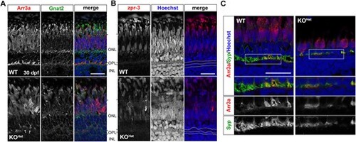
Photoreceptor synapses are disrupted in pomt1 KOHet retinae at 30 dpf. (A) Red/green double cone marker arrestin 3a (Arr3a) shows both disorganization in the outer segment and retraction of photoreceptors pedicles in the outer plexiform layer (OPL) (arrows). Cone-specific ?-transducin (Gnat2) staining is also reduced and lost in the outer limiting membrane. ONL: Outer nuclear layer; INL: Inner nuclear layer. Scale bar: 20 ?m B. Zpr-3 staining is reduced, but rods appear present in autofluorescence. Nuclear staining with Hoechst shows disorganization in the OPL and disruption in the nuclei of the horizontal cells below the OPL. OPL and horizontal cell nuclear layer are outlined by the white dotted lines. Scale bar: 20 ?m C. Synaptophysin (Syp) staining is discontinuous and showing loss of synaptic contacts. The white boxes outline the inset where immunostaining for each antibody is shown below. Scale bar: 20 ?m. Alt-text. This figure shows loss of photoreceptor synapses in pomt1 knock-out fish obtained from heterozygous mothers at 30 dpf.
|

