Fig. 1
- ID
- ZDB-FIG-240329-50
- Publication
- Endo et al., 2023 - Two zebrafish cacna1s loss-of-function variants provide models of mild and severe CACNA1S-related myopathy
- Other Figures
- All Figure Page
- Back to All Figure Page
|
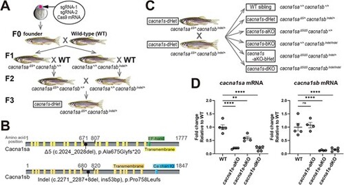
Generation of cacna1s mutant fish. (A) Schematic workflow of establishing cacna1s trans heterozygous mutants. sgRNA-1 was used for cacna1sa gene editing and sgRNA-2 for cacna1sb. The sequences of the gRNAs are described in the methods and Supplementary Fig. S1. (B) Schematics showing domains of Cacna1sa and Cacna1sb proteins and the obtained mutations. Yellow: transmembrane domain, Green: EF-hand domain, Blue (Ca chan IQ): voltage gated calcium channel IQ domain. (C) Progeny achieved by crossing cacna1s-dHet mutants. (D) mRNA levels of cacna1sa and cacna1sb in 6 dpf fish pools (WT; n = 5, aKO; n = 4, bKO; n = 4, dKO; n = 5). Values are Mean ± SEM (fold); cacna1sa: WT (1.00 ± 0.09), aKO (0.20 ± 0.01), bKO (0.59 ± 0.07), dKO (0.20 ± 0.04); cacna1sb: WT (1.00 ± 0.14), aKO (1.08 ± 0.10), bKO (0.13 ± 0.01), dKO (0.15 ± 0.03). Statistical analysis was performed using one-way ANOVA followed by Dunnett?s multiple comparisons test with P < 0.01 (**), P < 0.0001(****) or non-significance (ns). |

