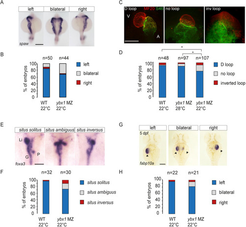
Mutant ybx1sa42 embryos have abnormal LR expression. (A) WISH to detect spaw in wild-type (n=50) and ybx1sa42 mutant (n=44) embryos at the 21-som stage. Scale bar: 100 µm. (B) Bar graph showing the proportion of embryos with spaw expression on the left (blue), bilaterally (grey) or on the right (red). Fisher's Exact two-tailed probability test P=0.0125. (C) Immunofluorescence showing MF20 labelling in the ventricle (red) and S46 labelling in the atrium (green) at 55 hpf. Representative D loop, no loop and inverted (inv) loop hearts are shown. A, atrium; V, ventricle. Scale bar: 100 µm. (D) Quantification of looping of the heart. Directional loop (D loop, blue), no loop (grey) and inverted looping (red) of the heart, with ybx1 mutant embryos at 22°C (n=107) showing a higher percentage of inverted and no loop hearts compared with control wild-type (WT; n=48) or ybx1 embryos at 28°C (n=97). An unpaired, two-tailed Student's t-test was used to analyse the data. *P<0.05. MZybx1 22°C compared with 28°C: P=0.022; MZybx1 22°C compared with WT: P=0.030. (E) WISH to detect foxa3 at 55 hpf in wild-type and ybx1 mutant embryos. G, gut; Li, liver; P, pancreas. Scale bar: 100 µm. (F) Quantification of visceral organ positioning in wild-type (n=32) and ybx1 mutant (n=30) embryos at 55 hpf showing an increase in situs ambiguus (grey) and situs inversus (red) with a decrease in situs solitus (blue) in ybx1 mutant embryos. Fisher's Exact two-tailed test. P=0.0113. (G) Expression of fabp10a at 5 dpf. Arrowheads indicate the liver. Scale bar: 100 µm. (H) Quantification of liver positioning in wild-type (n=22) and ybx1 mutant (n=21) embryos showing situs solitus (blue), situs ambiguus (grey) and situs inversus (red).
|
