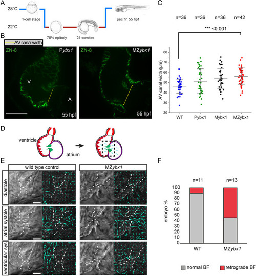
Ybx1 mutant embryos show abnormal heart morphogenesis. (A) Schematic of temperature shift experiments. ybx1 mutant embryos and controls were shifted to 22°C at 75% epiboly until 21-som and then grown at 28°C until 55 hpf. (B) Confocal images of zebrafish heart showing immunofluorescence with the ZN-8 antibody to label the of atrioventricular (AV) canal. Yellow arrows indicate the width of the AV canal in control Pybx1 and MZybx1 mutant embryos. A, atrium; V, ventricle. Scale bar: 25 µm. (C) Quantification AV canal width in µm from immunostaining of wild-type (WT; blue, n=36), Pybx1 (green, n=36), Mybx1 (black, n=36) and MZybx1 embryos (red, n=42). ANOVA test was used for statistical analysis (P=0.00016361; ***P<0.001). (D) Schematic of zebrafish heart at 5 dpf (left) and schematic of PIV analysis (right); black dashed box shows the area of the heart imaged and analysed. (E) Left: DIC images from movies of wild-type and ybx1 mutant hearts during diastole, atrial systole and ventricular systole. Right: PIV analysis of the images; cyan arrows indicate movements of red blood cells. Scale bars: 20 µm. (F) Percentage of embryos showing normal blood flow (BF) versus retrograde BF in wild-type (n=11) and ybx1 mutant (n=12) embryo groups.
|
