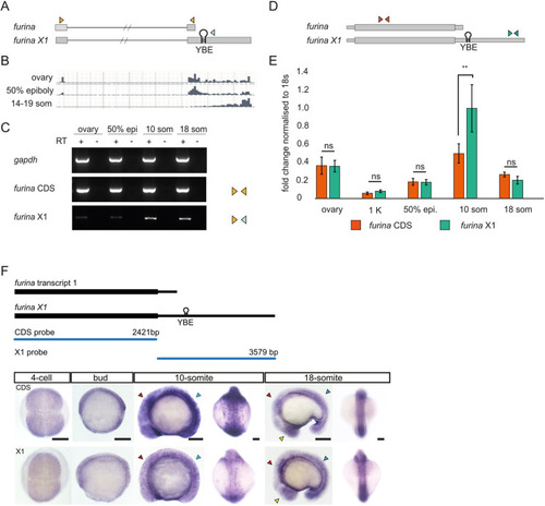
Zebrafish furina transcripts show different 3′UTR lengths, with a predominant long isoform expressed during somitogenesis. (A) Schematic of furina transcript 1 and variant X1. Orange arrowheads represent primers to amplify transcript 1; one orange and one green primer were used to detect variant X1. Hairpin indicates the YBE motif in the 3′UTR of X1. (B) RNA sequencing excerpt from ovary, 50% epiboly and 14-19 som stages. (C) Semi-quantitative RT-PCR detecting gapdh positive control, furina coding sequences (CDS) and furina variant X1 3′UTR (X1). (D) Schematic of primers used in qPCR to detect the furina CDS and variant X1. The orange primer set was used to amplify the CDS and green primers used to amplify variant X1. (E) Quantitative PCR analysis from ovary, 1 K, 50% epiboly, 10-som and 18-som embryos. Orange bars represent furina coding region, green bars show exclusively variant X1. **P<0.01 in unpaired, two-tailed Student's t-test. n=50 embryos per stage for mRNA extraction, P=0.014. ns, not significant. (F) Whole-mount in situ hybridisation with probes that detect the CDS or variant X1. Yellow arrowheads point to enrichment in eyes, red in hindbrain and blue in somites. Lateral and dorsal views are shown at 10-som and 18-som. Scale bars: 100 µm.
|
