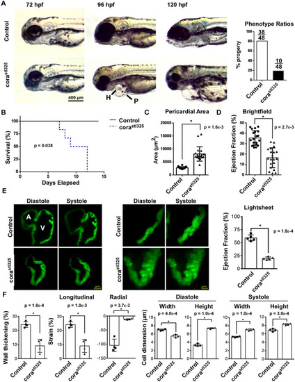
corastl325/stl325 embryos display reduced survival and heart failure. (A) Light microscopy showing the emergence of pericardial edema between 72 and 120 h post fertilization (hpf). P, pericardium; H, heart. Phenotype ratios are presented on the right. (number of trials=4; number of samples per trial=48) (B) Survival curve of corastl325 embryos relative to their unaffected clutchmates (number of trials=4; number of samples per trial=48). (C) Quantification of light microscopy images measuring area of the pericardium (number of trials=4; number of samples per trial=48). (D) Quantification of light microscopy images measuring ejection fraction (number of trials=4; number of samples per trial=48). (E) Lightsheet microscopy generated images of 96 hpf embryos harboring the cmlc::GFP transgenic reporter. Hearts are shown in systole and diastole. A, atrium; V, ventricle. Quantification of ejection fraction (n=4). (F) Quantification of changes in cell dimension using lightsheet-generated videos (n=4).
|
