
Fig 4
- ID
- ZDB-FIG-221121-11
- Publication
- Sarmah et al., 2022 - Elf3 deficiency during zebrafish development alters extracellular matrix organization and disrupts tissue morphogenesis
- Other Figures
- All Figure Page
- Back to All Figure Page
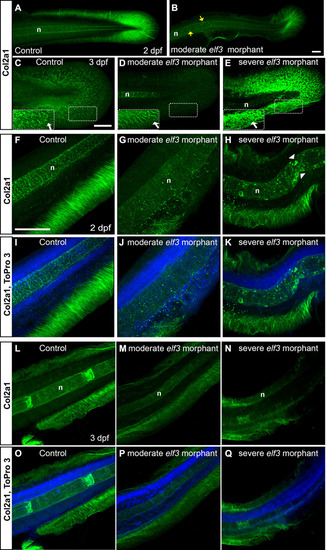
Elf3 deficiency led to disruption of Col2α1 expression in the fin and notochord.
(A-E) 3D reconstruction of the confocal images of the Col2α1 antibody stained embryos show dense Col2α1 fibrils radially distributed throughout the whole caudal fin in the control (A, C) and less dense, irregularly distributed fibrils in moderately affected morphants (B, D); severely affected morphant showed abnormal accumulation of Col2α1, but no fibrillary pattern (E). Insets show high magnification of the marked rectangle area. yellow arrow: abnormal Col2α1 accumulation within notochord; white arrow: Col2α1 fibrils. (F-Q) Optical section of the confocal images of the Col2α1 antibody stained embryos show severely disrupted and disorganization median fin fold collagen fibrils in the elf3 morphants at 2 dpf (G, H, J, K) and 3 dpf larva (M, N, P, Q) and nicely organized, thick fibrils in the control (F, I, L, O). Scale bar for A, B = 50 μm; for C-E = 100 μm; and for F-Q = 100 μm. |
| Fish: | |
|---|---|
| Knockdown Reagents: | |
| Observed In: | |
| Stage Range: | Long-pec to Protruding-mouth |